Synucleinopathies are neurological diseases that are characterized by the accumulation of aggregates of a cytosolic protein, α-synuclein, at the plasma membrane. Even though the pathological role of the protein is established, the mechanism by which it damages neurons remains unclear due to the difficulty to correctly mimic the plasma membrane in vitro. Using a microfluidic setup in which the composition of the plasma membrane, including the asymmetry of the two leaflets, is recapitulated, we demonstrate a triple action of α-synuclein on the membrane. First, it changes membrane topology by inducing pores of discrete sizes, likely nucleated from membrane-bound proteins and subsequently enlarged by proteins in solution. Second, protein binding to the cytosolic leaflet increases the membrane capacitance by thinning it and/or changing its relative permittivity. Third, α-synuclein insertion inside the membrane hydrophobic core immobilizes the lipids in both leaflets, including the opposing protein-free extracellular one.
Publis
JUNO, the receptor of sperm IZUMO1, is expressed by the human oocyte and is essential for human fertilisation
STUDY QUESTION: Is JUNO protein present at the surface membrane of human oocytes and involved in the fertilisation process?
SUMMARY ANSWER: JUNO protein is expressed on the plasma membrane of human oocytes and its inhibition by a monoclonal antibody completely blocks gamete fusion.
WHAT IS KNOWN ALREADY: Fusion of gamete membranes is the culminating event of the fertilisation process, but its molecular mechanisms are poorly understood. Until now, three molecules have been shown to be essential: CD9 tetraspanin in the oocyte, Izumo1 protein on the sperm and Juno, its corresponding receptor on the oocyte. Oocyte CD9 and sperm IZUMO1 have been identified in human gametes and their interaction is also well-conserved among several mammalian species. The presence of JUNO on human oocytes, however, has not yet been reported, nor has its role in fertilisation been investigated.
STUDY DESIGN, SIZE, DURATION: We selected an anti-human JUNO antibody in order to investigate the presence of JUNO on the oocyte membrane surface and studied its potential involvement in gamete membrane interaction during fertilisation.
PARTICIPANTS/MATERIALS, SETTING, METHODS: Monoclonal antibodies against human JUNO (anti-hJUNO mAb) were produced by immunisation of mice with HEK cells transfected with the putative human JUNO sequence (HEK-hJUNO). These antibodies were used for immunostaining experiments and in vitro fertilisation assays with human gametes (GERMETHEQUE Biobank).
MAIN RESULTS AND THE ROLE OF CHANCE: Three hybridoma supernatants, verified by immunostaining, revealed specifically HEK-hJUNO cells. The three purified monoclonal antibodies, FJ2E4 (IgG1), FJ8E8 (IgG1) and FJ4F5 (IgG2a), recognised the soluble recombinant human JUNO protein and, in a western blot of HEK-hJUNO extracts, a protein with an expected MW of 25 kDa. In addition, soluble recombinant human IZUMO protein inhibited the binding of anti-hJUNO mAbs to cells expressing hJUNO. Using these anti-hJUNO mAbs in immunostaining, we identified the presence of JUNO protein at the plasma membrane of human oocytes. Furthermore, we revealed a progressive expression of JUNO according to oocyte maturity. Finally, we showed that human zona-free oocytes, inseminated in the presence of anti-hJUNO mAb, were not fertilised by human sperm. These results suggest that, as seen in the mouse, JUNO is indeed involved in human gamete membrane fusion during fertilisation.
Anisotropic cellular forces support mechanical integrity of the Stratum Corneum barrier
The protective function of biological surfaces that are exposed to the exterior of living organisms is the result of a complex arrangement and interaction of cellular components. This is the case for the most external cornified layer of skin, the stratum corneum (SC). This layer is made of corneocytes, the elementary ‘flat bricks’ that are held together through adhesive junctions. Despite the well-known protective role of the SC under high mechanical stresses and rapid cell turnover, the subtleties regarding the adhesion and mechanical interaction among the individual corneocytes are still poorly known. Here, we explore the adhesion of single corneocytes at different depths of the SC, by pulling them using glass microcantilevers, and measuring their detachment forces. We measured their interplanar adhesion between SC layers, and their peripheral adhesion among cells within a SC layer. Both adhesions increased considerably with depth. At the SC surface, with respect to adhesion, the corneocyte population exhibited a strong heterogeneity, where detachment forces differed by more than one order of magnitude for corneocytes located side by side. The measured detachment forces indicated that in the upper-middle layers of SC, the peripheral adhesion was stronger than the interplanar one. We conclude that the stronger peripheral adhesion of corneocytes in the SC favors an efficient barrier which would be able to resist strong stresses.
Highly Reproducible Physiological Asymmetric Membrane with Freely Diffusing Embedded Proteins in a 3D-Printed Microfluidic Setup
Experimental setups to produce and to monitor model membranes have been successfully used for decades and brought invaluable insights into many areas of biology. However, they all have limitations that prevent the full in vitro mimicking and monitoring of most biological processes. Here, a suspended physiological bilayer-forming chip is designed from 3D-printing techniques. This chip can be simultaneously integrated to a confocal microscope and a path-clamp amplifier. It is composed of poly(dimethylsiloxane) and consists of a ≈100 μm hole, where the horizontal planar bilayer is formed, connecting two open crossed-channels, which allows for altering of each lipid monolayer separately. The bilayer, formed by the zipping of two lipid leaflets, is free-standing, horizontal, stable, fluid, solvent-free, and flat with the 14 types of physiologically relevant lipids, and the bilayer formation process is highly reproducible. Because of the two channels, asymmetric bilayers can be formed by making the two lipid leaflets of different composition. Furthermore, proteins, such as transmembrane, peripheral, and pore-forming proteins, can be added to the bilayer in controlled orientation and keep their native mobility and activity. These features allow in vitro recapitulation of membrane process close to physiological conditions.
SNARE machinery is optimized for ultrafast fusion
SNARE proteins zipper to form complexes (SNAREpins) that power vesicle fusion with target membranes in a variety of biological processes. A single SNAREpin takes about 1 s to fuse two bilayers, yet a handful can ensure release of neurotransmitters from synaptic vesicles much faster: in a 10th of a millisecond. We propose that, similar to the case of muscle myosins, the ultrafast fusion results from cooperative action of many SNAREpins. The coupling originates from mechanical interactions induced by confining scaffolds. Each SNAREpin is known to have enough energy to overcome
the fusion barrier of 25–35 kBT; however, the fusion barrier only becomes relevant when the SNAREpins are nearly completely zippered, and from this state, each SNAREpin can deliver only a small fraction of this energy as mechanical work. Therefore, they have to act cooperatively, and we show that at least three of them are needed to ensure fusion in less than a millisecond. However, to reach the prefusion state collectively, starting from
the experimentally observed half-zippered metastable state, the SNAREpins have to mechanically synchronize, which takes more time as the number of SNAREpins increases. Incorporating this somewhat counterintuitive idea in a simple coarse-grained model results in the prediction that there should be an optimum number of SNAREpins for submillisecond fusion: three to six over a wide range of parameters. Interestingly, in situ cryoelectron microscope tomography has very recently shown that exactly six SNAREpins participate in the fusion of each synaptic vesicle. This number is in the range predicted by our theory.
Synaptotagmin oligomers are necessary and can be sufficient to form a Ca2+ sensitive fusion clamp
The buttressed-ring hypothesis, supported by recent cryo-electron tomography analysis of docked synaptic-like vesicles in neuroendocrine cells, postulates that prefusion SNAREpins are stabilized and organized by Synaptotagmin (Syt) ring-like oligomers. Here, we use a reconstituted single-vesicle fusion analysis to test the prediction that destabilizing the Syt1 oligomers destabilizes the clamp and results in spontaneous fusion in the absence of Ca2+. Vesicles in which Syt oligomerization is compromised by a ring-destabilizing mutation dock and diffuse freely on the bilayer until they fuse spontaneously, similar to vesicles containing only v-SNAREs. In contrast, vesicles containing wild-type Syt are immobile as soon as they attach to the bilayer and remain frozen in place, up to at least 1 h until fusion is triggered by Ca2+.
S-Palmitoylation Sorts Membrane Cargo for Anterograde Transport in the Golgi
While retrograde cargo selection in the Golgi is known to depend on specific signals, it is unknown whether anterograde cargo is sorted, and anterograde signals have not been identified. We suggest here that S-palmitoylation of anterograde cargo at the Golgi membrane interface is an anterograde signal and that it results in concentration in curved regions at the Golgi rims by simple physical chemistry. The rate of transport across the Golgi of two S-palmitoylated membrane proteins is controlled by S-palmitoylation. The bulk of S-palmitoylated proteins in the Golgi behave analogously, as revealed by click chemistry-based fluorescence and electron microscopy. These palmitoylated cargos concentrate in the most highly curved regions of the Golgi membranes, including the fenestrated perimeters of cisternae and associated vesicles. A palmitoylated transmembrane domain behaves similarly in model systems.
High-Throughput Monitoring of Single Vesicle Fusion Using Freestanding Membranes and Automated Analysis
In vivo membrane fusion primarily occurs between highly curved vesicles and planar membranes. A better understanding of fusion entails an accurate in vitro reproduction of the process. To date, supported bilayers have been commonly used to mimic the planar membranes. Soluble
N-ethylmaleimide-sensitive factor attachment protein receptor (SNARE) proteins that induce membrane fusion usually have limited fluidity when embedded in supported bilayers. This alters the kinetics and prevents correct reconstitution of the overall fusion process. Also, observing content release across the membrane is hindered by the lack of a second aqueous compartment. Recently, a step toward resolving these issues was achieved by using membranes spread on holey substrates. The mobility of proteins was preserved but vesicles were prone to bind to the substrate when reaching the edge of the hole, preventing the observation of many fusion events over the suspended membrane. Building on this recent advance, we designed a method for the formation of pore-spanning lipid bilayers containing t-SNARE proteins on Si/SiO2 holey chips, allowing the observation of many individual vesicle fusion events by both lipid mixing and content release. With this setup, proteins embedded in the suspended membrane bounced back when they reached the edge of the hole which ensured vesicles did not bind to the substrate. We observed SNARE-dependent membrane fusion with the freestanding bilayer of about 500 vesicles. The time between vesicle docking and fusion is ∼1 s. We also present a new multimodal open-source software, Fusion Analyzer Software, which is required for fast data analysis.
Vesicle Tubulation with Self-Assembling DNA Nanosprings
A major goal of nanotechnology and bioengineering is to build artificial nanomachines capable of generating specific membrane curvatures on demand. Inspired by natural membrane-deforming proteins, we designed DNA-origami curls that polymerize into nanosprings and show their efficacy in vesicle deformation. DNA-coated membrane tubules emerge from spherical vesicles when DNA-origami polymerization or high membrane-surface coverage occurs. Unlike many previous methods, the DNA self-assembly-mediated membrane tubulation eliminates the need for detergents or topdown manipulation. The DNA-origami design and deformation conditions have substantial influence on the tubulation efficiency and tube morphology, underscoring the intricate interplay between lipid bilayers and vesicle-deforming DNA structures.
Rearrangements under confinement lead to increased binding energy of Synaptotagmin-1 with anionic membranes in Mg2+ and Ca2+
Synaptotagmin-1 (Syt1) is the primary calcium sensor (Ca2+) that mediates neurotransmitter release at the synapse. The tandem C2 domains (C2A and C2B) of Syt1 exhibit functionally critical, Ca2+-dependent interactions with the plasma membrane. With the surface forces apparatus, we directly measure the binding energy of membrane-anchored Syt1 to an anionic membrane and find that Syt1 binds with ~6 kBT in EGTA, ~10 kBT in Mg2+ and ~18 kBT in Ca2+. Molecular rearrangements measured during confinement are more prevalent in Ca2+ and Mg2+ and suggest that Syt1 initially binds through C2B, then reorients the C2 domains into the preferred binding configuration. These results provide energetic and mechanistic details of the Syt1 Ca2+-activation process in synaptic transmission.
Egg CD9 protein tides correlated with sperm oscillations tune the gamete fusion ability in mammal
Mammalian fertilization involves membrane events -adhesion, fusion, sperm engulfment, membrane block to polyspermy- whose causes remain largely unknown. Recently, specific oscillations of the sperm in contact with the egg were shown to be necessary for fusion. Using a microfluidic chip to impose the venue for the encounter of two gametes allowed real-time observation of the membrane remodelling occurring at the sperm/egg interface. The spatiotemporal mapping of egg CD9 revealed that this protein concentrates at the egg/sperm interface as a result of sperm oscillations, until a CD9-rich platform is nucleated on which fusion immediately takes place. Within 2 to 5 minutes after fusion, most of the CD9 leaves the egg for the external aqueous medium. Then an egg membrane wave engulfs the sperm head in approximately 25 minutes. These results show that sperm oscillations initiate the CD9 recruitment that causes gamete fusion after which CD9 and associated proteins leave the membrane in a process likely to contribute to block polyspermy. They highlight that the gamete fusion story in mammals is an unexpected interplay between mechanical constraints and proteins.
Hypothesis – buttressed rings assemble, clamp, and release SNAREpins for synaptic transmission
Neural networks are optimized to detect temporal coincidence on the millisecond timescale. Here, we offer a synthetic hypothesis based on recent structural insights into SNAREs and the C2 domain proteins to explain how synaptic transmission can keep this pace. We suggest that an outer ring of up to six curved Munc13 ‘MUN’ domains transiently anchored to the plasma membrane via its flanking domains surrounds a stable inner ring comprised of synaptotagmin C2 domains to serve as a work-bench on which SNAREpins are templated. This ‘buttressed-ring hypothesis’ affords straightforward answers to many principal and long-standing questions concerning how SNAREpins can be assembled, clamped, and then released synchronously with an action potential.
Circular oligomerization is an intrinsic property of synaptotagmin
Previously, we showed that synaptotagmin1 (Syt1) forms Ca2+-sensitive ring-like oligomers on membranes containing acidic lipids and proposed a potential role in regulating neurotransmitter release. Here, we report that Syt1 assembles into similar ring-like oligomers in solution when triggered by naturally occurring polyphosphates (PIP2 and ATP) and magnesium ions (Mg2+). These soluble Syt1 rings were observed by electron microscopy and independently demonstrated and quantified using fluorescence correlation spectroscopy. Oligomerization is triggered when polyphosphates bind to the polylysine patch in C2B domain and is stabilized by Mg2+, which neutralizes the Ca2+-binding aspartic acids that likely contribute to the C2B interface in the oligomer. Overall, our data show that ring-like polymerization is an intrinsic property of Syt1 with reasonable affinity that can be triggered by the vesicle docking C2B-PIP2 interaction and raise the possibility that Syt1 rings could pre-form on the synaptic vesicle to facilitate docking.
Placing and shaping liposomes with reconfigurable DNA nanocages
The diverse structure and regulated deformation of lipid bilayer membranes are among a cell’s most fascinating features.
Artificial membrane-bound vesicles, known as liposomes, are versatile tools for modelling biological membranes and
delivering foreign objects to cells. To fully mimic the complexity of cell membranes and optimize the efficiency of delivery
vesicles, controlling liposome shape (both statically and dynamically) is of utmost importance. Here we report the
assembly, arrangement and remodelling of liposomes with designer geometry: all of which are exquisitely controlled by a
set of modular, reconfigurable DNA nanocages. Tubular and toroid shapes, among others, are transcribed from DNA cages
to liposomes with high fidelity, giving rise to membrane curvatures present in cells yet previously difficult to construct in
vitro. Moreover, the conformational changes of DNA cages drive membrane fusion and bending with predictable outcomes,
opening up opportunities for the systematic study of membrane mechanics.
Axon tension regulates fasciculation/ defasciculation through the control of axon shaft zippering
While axon fasciculation plays a key role in the development of neural networks, very
little is known about its dynamics and the underlying biophysical mechanisms. In a model system
composed of neurons grown ex vivo from explants of embryonic mouse olfactory epithelia, we
observed that axons dynamically interact with each other through their shafts, leading to zippering
and unzippering behavior that regulates their fasciculation. Taking advantage of this new
preparation suitable for studying such interactions, we carried out a detailed biophysical analysis of
zippering, occurring either spontaneously or induced by micromanipulations and pharmacological
treatments. We show that zippering arises from the competition of axon-axon adhesion and
mechanical tension in the axons, and provide the first quantification of the force of axon-axon
adhesion. Furthermore, we introduce a biophysical model of the zippering dynamics, and we
quantitatively relate the individual zipper properties to global characteristics of the developing
axon network. Our study uncovers a new role of mechanical tension in neural development: the
regulation of axon fasciculation.
Actual fusion efficiency in the lipid mixing assay – Comparison between nanodiscs and liposomes
Lipid exchange occurs between membranes during fusion or active lipid transfer. These processes are necessary in vivo for the homeostasis of the cell at the level of the membranes, the organelles and the cell itself. They are also used by the cell to interact with the surrounding medium. Several assays have been developed to characterize in vitro these processes on model systems. The most common one, relying on fluorescence dequenching, measures lipid mixing between small membranes such as liposomes or nanodiscs in bulk. Usually, relative comparisons of the rate of lipid exchange are made between measurements performed in parallel. Here, we establish a quantitative standardization of this assay to avoid any bias resulting from the temperatures, the chosen fluorescent lipid fractions and from the various detergents used to normalize the measurements. We used this standardization to quantitatively compare the efficiency of SNARE-induced fusion in liposome-liposome and liposome-nanodisc configurations having similar collision frequency. We found that the initial yield of fusion is comparable in both cases, 1 per 2–3 million collisions in spite of a much larger dequenching signal with nanodiscs. Also, the long-term actual fusion rate is slightly lower with nanodiscs than in the liposome-liposome assay.
BFPTool: a software tool for analysis of Biomembrane Force Probe experiments
Background: The Biomembrane Force Probe is an approachable experimental technique commonly used for single-molecule force spectroscopy and experiments on biological interfaces. The technique operates in the range of forces from 0.1 pN to 1000 pN. Experiments are typically repeated many times, conditions are often not optimal, the captured video can be unstable and lose focus; this makes efficient analysis challenging, while out-of-the-box non-proprietary solutions are not freely available.
Results: This dedicated tool was developed to integrate and simplify the image processing and analysis of
videomicroscopy recordings from BFP experiments. A novel processing feature, allowing the tracking of the pipette, was incorporated to address a limitation of preceding methods. Emphasis was placed on versatility and comprehensible user interface implemented in a graphical form.
Conclusions: An integrated analytical tool was implemented to provide a faster, simpler and more convenient way to process and analyse BFP experiments.
Low energy cost for optimal speed and control of membrane fusion
Membrane fusion is the cell’s delivery process, enabling its many compartments to receive cargo and machinery for cell growth and intercellular communication. The overall activation energy of the process must be large enough to prevent frequent and nonspecific spontaneous fusion events, yet must be low enough to allow it to be overcome upon demand by specific fusion proteins [such as soluble N-ethylmaleimide–sensitive factor attachment protein receptors (SNAREs)]. Remarkably, to the best of our knowledge, the activation energy for spontaneous bilayer fusion has never been measured. Multiple models have been developed and refined to estimate the overall activation energy and its component parts, and they span a very broad range from 20 kBT to 150 kBT, depending on the assumptions. In this study, using a bulk lipid-mixing assay at various temperatures, we report that the activation energy of complete membrane fusion is at the lowest range of these theoretical values. Typical lipid vesicles were found to slowly and spontaneously fully fuse with activation energies of ∼30 kBT. Our data demonstrate that the merging of membranes is not nearly as energy consuming as anticipated by many models and is ideally positioned to minimize spontaneous fusion while enabling rapid, SNARE-dependent fusion upon demand.
Endothelial basement membrane laminin 511 is essential for shear stress response
Abstract:
Shear detection and mechanotransduction by arterial endothelium requires junctional complexes containing PECAM-1 and VE-cadherin, as well as firm anchorage to the underlying basement membrane. While considerable information is available for junctional complexes in these processes, gained largely from in vitro studies, little is known about the contribution of the endothelial basement membrane. Using resistance artery explants, we show that the integral endothelial basement membrane component, laminin 511 (laminin a5), is central to shear detection and mechanotransduction and its elimination at this site results in ablation of dilation in response to increased shear stress. Loss of endothelial laminin 511 correlates with reduced cortical stiffness of arterial endothelium in vivo, smaller integrin b1-positive/vinculin-positive focal adhesions, and reduced junctional association of actin–myosin II. In vitro assays reveal that b1 integrin-mediated interaction with laminin 511 results in high strengths of adhesion, which promotes p120 catenin association with VE-cadherin, stabilizing it at cell junctions and increasing cell–cell adhesion strength. This highlights the importance of endothelial laminin 511 in shear response in the physiologically relevant context of resistance arteries.
Stability, folding dynamics, and long-range conformational transition of the synaptic t-SNARE complex
Abstract:
Synaptic soluble N-ethylmaleimide–sensitive factor attachment protein receptors (SNAREs) couple their stepwise folding to fusion of synaptic vesicles with plasma membranes. In this process, three SNAREs assemble into a stable four-helix bundle. Arguably, the first and rate-limiting step of SNARE assembly is the formation of an activated binary target (t)-SNARE complex on the target plasma membrane, which then zippers with the vesicle (v)-SNARE on the vesicle to drive membrane fusion. However, the t-SNARE complex readily misfolds, and its structure, stability, and dynamics are elusive. Using single-molecule force spectroscopy, we modeled the synaptic t-SNARE complex as a parallel three-helix bundle with a small frayed C terminus. The helical bundle sequentially folded in an N-terminal domain (NTD) and a C-terminal domain (CTD) separated by a central ionic layer, with total unfolding energy of ∼17 kBT, where kB is the Boltzmann constant
and T is 300 K. Peptide binding to the CTD activated the t-SNARE complex to initiate NTD zippering with the v-SNARE, a mechanism likely shared by the mammalian uncoordinated-18-1 protein (Munc18-1). The NTD zippering then dramatically stabilized the CTD, facilitating further SNARE zippering. The subtle bidirectional t-SNARE conformational switch was mediated by the ionic layer. Thus, the t-SNARE complex acted as a switch to enable fast and controlled SNARE zippering required for synaptic vesicle fusion and neurotransmission.
A specific flagellum beating mode for inducing fusion in mammalian fertilization and kinetics of sperm internalization
The salient phases of fertilization are gamete adhesion, membrane fusion, and internalization of the spermatozoon into the oocyte but the precise timeline and the molecular, membrane and cell mechanisms underlying these highly dynamical events are far from being established. The high motility of the spermatozoa and the unpredictable location of sperm/egg fusion dramatically hinder the use of real time imaging optical techniques that should directly provide the dynamics of cell events. Using an approach based on microfluidics technology, the sperm/egg interaction zone was imaged with the best front view, and the timeline of the fertilization events was established with an unparalleled temporal accuracy from the onset of gamete contact to full sperm DNA decondensation. It reveals that a key element of the adhesion phase to initiate fusion is the oscillatory motion of the sperm head on the oocyte plasma membrane generated by a specific flagellum-beating mode. It also shows that the incorporation of the spermatozoon head is a two steps process that includes simultaneous diving, tilt, and plasma membrane degradation of the sperm head into the oocyte and subsequent DNA decondensation.
FRAP to Characterize Molecular Diffusion and Interaction in Various Membrane Environments
Fluorescence recovery after photobleaching (FRAP) is a standard method used to study the dynamics of lipids and proteins in artificial and cellular membrane systems. The advent of confocal microscopy two decades ago has made quantitative FRAP easily available to most laboratories. Usually, a single bleaching pattern/area is used and the corresponding recovery time is assumed to directly provide a diffusion coefficient, although this is only true in the case of unrestricted Brownian motion. Here, we propose some general guidelines to perform FRAP experiments under a confocal microscope with different bleaching patterns and area, allowing the experimentalist to establish whether the molecules undergo Brownian motion (free diffusion) or whether they have restricted or directed movements. Using in silico simulations of FRAP measurements, we further indicate the data acquisition criteria that have to be verified in order to obtain accurate values for the diffusion coefficient and to be able to distinguish between different diffusive species. Using this approach, we compare the behavior of lipids in three different membrane platforms (supported lipid bilayers, giant liposomes and sponge phases), and we demonstrate that FRAP measurements are consistent with results obtained using other techniques such as Fluorescence Correlation Spectroscopy (FCS) or Single Particle Tracking (SPT). Finally, we apply this method to show that the presence of the synaptic protein Munc18-1 inhibits the interaction between the synaptic vesicle SNARE protein, VAMP2, and its partner from the plasma membrane, Syn1A.
On-Chip Quantitative Measurement of Mechanical Stresses During Cell Migration with Emulsion Droplets
Abstract:
The ability of immune cells to migrate within narrow and crowded spaces is a critical feature involved in various physiological processes from immune response to metastasis. Several in-vitro techniques have been developed so far to study the behaviour of migrating cells, the most recent being based on the fabrication of microchannels within which cells move. To address the question of the mechanical stress a cell is able to produce during the encounter of an obstacle while migrating, we developed a hybrid microchip made of parallel PDMS channels in which oil droplets are sparsely distributed and serve as deformable obstacles. We thus show that cells strongly deform droplets while passing them. Then, we show that the microdevice can be used to study the influence of drugs on migration at the population level. Finally, we describe a quantitative analysis method of the droplet deformation that allows measuring in real-time the mechanical stress exerted by a single cell. The method presented herein thus constitutes a powerful analytical tool for cell migration studies under confinement.
Control of plasma membrane lipid homeostasis by the extended synaptotagmins
Abstract:
Acute metabolic changes in plasma membrane (PM) lipids, such as those mediating signalling reactions, are rapidly compensated by homeostatic responses whose molecular basis is poorly understood. Here we show that the extended synaptotagmins (E-Syts), endoplasmic reticulum (ER) proteins that function as PtdIns(4,5)P2- and Ca2C-regulated tethers to the PM, participate in these responses. E-Syts transfer glycerolipids between bilayers in vitro, and this transfer requires Ca2C and their lipid-harbouring SMP domain. Genome-edited cells lacking E-Syts do not exhibit abnormalities in the major glycerolipids at rest, but exhibit enhanced and sustained accumulation of PM diacylglycerol following PtdIns(4,5)P2 hydrolysis by PLC activation, which can be rescued by expression of E-Syt1, but not by mutant E-Syt1 lacking the SMP domain. The formation of E-Syt-dependent ER–PM tethers in response to stimuli that cleave PtdIns(4,5)P2 and elevate Ca2C may help reverse accumulation of diacylglycerol in the PM by transferring it to the ER for metabolic recycling.
Snapshot of sequential SNARE assembling states between membranes shows that N-terminal transient assembly initializes fusion
Abstract:
Many prominent biological processes are driven by protein assembling between membranes. Understanding the mechanisms then entails determining the assembling pathway of the involved proteins. Because the intermediates are by nature transient and located in the intermembrane space, this determination is generally a very difficult, not to say intractable, problem. Here, by designing a setup with sphere/plane geometry, we have been able to freeze one transient state in which the N-terminal domains of SNARE proteins are assembled. A single camera frame is sufficient to obtain the complete probability of this state with the transmembrane distance. We show that it forms when membranes are 20 nm apart and stabilizes by further assembling of the SNAREs at 8 nm. This setup that fixes the intermembrane distance, and thereby the transient states, while optically probing the level of molecular assembly by Förster resonance energy transfer (FRET) can be used to characterize any other transient transmembrane complexes.
Kinetic barriers to SNAREpin assembly in the regulation of membrane docking/priming and fusion
Abstract:
Neurotransmission is achieved by soluble NSF attachment protein receptor (SNARE)-driven fusion of readily releasable vesicles that are docked and primed at the presynaptic plasma membrane. After neurotransmission, the readily releasable pool of vesicles must be refilled in less than 100 ms for subsequent release. Here we show that the initial association of SNARE complexes, SNAREpins, is far too slow to support this rapid refilling owing to an inherently high activation energy barrier. Our data suggest that acceleration of this process, i.e., lowering of the barrier, is physiologically necessary and can be achieved by molecular factors. Furthermore, under zero force, a low second energy barrier transiently traps SNAREpins in a half-zippered state similar to the partial assembly that engages calcium-sensitive regulatory machinery. This result suggests that the barrier must be actively raised in vivo to generate a sufficient pause in the zippering process for the regulators to set in place. We show that the heights of the activation energy barriers can be selectively changed by molecular factors. Thus, it is possible to modify, both in vitro and in vivo, the lifespan of each metastable state. This controllability provides a simple model in which vesicle docking/priming, an intrinsically slow process, can be substantially accelerated. It also explains how the machinery that regulates vesicle fusion can be set in place while SNAREpins are trapped in a halfzippered state.
A Programmable DNA Origami Platform to Organize SNAREs for Membrane Fusion
Soluble N-ethylmaleimide-sensitive factor attachment protein receptor (SNARE) complexes are the core molecular machinery of membrane fusion, a fundamental process that drives inter- and intracellular communication and trafficking. One of the questions that remains controversial has been whether and how SNAREs cooperate. Here we show the use of self-assembled DNA-nanostructure rings to template uniform-sized small unilamellar vesicles containing predetermined maximal number of externally facing SNAREs to study the membrane-fusion process. We also incorporated lipid-conjugated complementary ssDNA as tethers into vesicle and target membranes, which enabled bypass of the rate-limiting docking step of fusion reactions and allowed direct observation of individual membrane-fusion events at SNARE densities as low as one pair per vesicle. With this platform, we confirmed at the single event level that, after docking of the templated-SUVs to supported lipid bilayers (SBL), one to two pairs of SNAREs are sufficient to drive fast lipid mixing. Modularity and programmability of this platform makes it readily amenable to studying more complicated systems where auxiliary proteins are involved.
Accelerating SNARE Mediated Membrane Fusion by DNA-Lipid Tethers
SNARE proteins are the core machinery to drive fusion of a vesicle with its target membrane. Inspired by the tethering proteins that bridge the membranes and thus prepare SNAREs for docking and fusion, we developed a lipid-conjugated ssDNA mimic that is capable of regulating SNARE function, in situ. The DNA-lipid tethers consist of a 21 base pairs binding segment at the membrane distal end that can bridge two liposomes via specific base-pair hybridization. A linker at the membrane proximal end is used to control the separation distance between the liposomes. In the presence of these artificial tethers, SNARE-mediated lipid mixing is significantly accelerated, and the maximum fusion rate is obtained with the linker shorter than 40 nucleotides. As a programmable tool orthogonal to any native proteins, the DNA-lipid tethers can be further applied to regulate other biological processes where capturing and bridging of two membranes are the prerequisites for the subsequent protein function.
The Energy of COPI for Budding Membranes
As a major actor of cellular trafficking, COPI coat proteins assemble on membranes and locally bend them to bud 60 nm-size coated particles. Budding requires the energy of the coat assembly to overcome the one necessary to deform the membrane which primarily depends on the bending modulus and surface tension, γ. Using a COPI-induced oil nano-droplet formation approach, we modulated the budding of nanodroplets using various amounts and types of surfactant. We found a Heaviside-like dependence between the budding efficiency and γ : budding was only dependent on γ and occurred beneath 1.3 mN/m. With the sole contribution of γ to the membrane deformation energy, we assessed that COPI supplies ~1500 kBT for budding particles from membranes, which is consistent with common membrane deformation energies. Our results highlight how a simple remodeling of the composition of membranes could mechanically modulate budding in cells.
Stability of C(12)E(j) Bilayers Probed with Adhesive Droplets.
The stability of model surfactant bilayers from the poly(ethylene glycol) mono-n-dodecyl ether (C12Ej) family was probed. The surfactant bilayers were formed by the adhesion of emulsion droplets. We generated C12Ej bilayers by forming water-in-oil (w/o) emulsions with saline water droplets, covered by the surfactant, in a silicone and octane oil mixture. Using microfluidics, we studied the stability of those bilayers. C12E1 allowed only short-lived bilayers whereas C12E2 bilayers were stable over a wide range of oil mixtures. At high C12E2 concentration, a two-phase region was displayed in the phase diagram: bilayers formed by the adhesion of two water droplets and Janus-like particles consisting of adhering aqueous and amphiphilic droplets. C12E8 and C12E25 did not mediate bilayer formation and caused phase inversion leading to o/w emulsion. With intermediate C12E4 and C12E5 surfactants, both w/o and o/w emulsions were unstable. We provided the titration of the C12E2 bilayer with C12E4 and C12E5 to study and predict their stability behavior.
Formation of Giant Unilamellar Proteo-Liposomes by Osmotic Shock
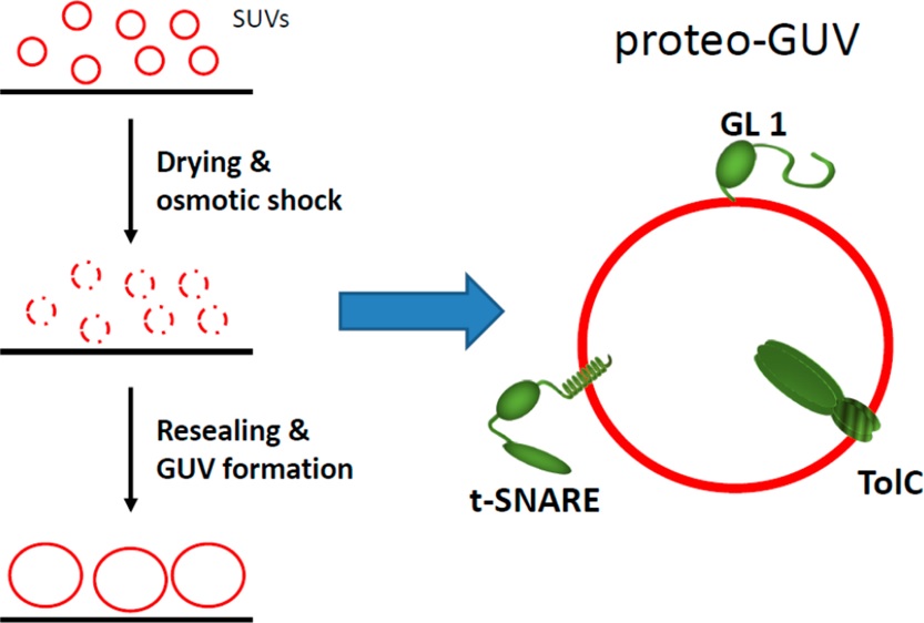
Giant unilamellar vesicles (GUVs), composed of a phospholipid bilayer, are often used as a model system for cell membranes. However, the study of proteo-membrane interactions in this system is limited as the incorporation of integral and lipid-anchored proteins into GUVs remains challenging. Here, we present a simple generic method to incorporate proteins into GUVs. The basic principle is to break proteo-liposomes with an osmotic shock. They subsequently reseal into larger vesicles which, if necessary, can endure the same to obtain even larger proteo-GUVs. This process does not require specific lipids or reagents, works under physiological conditions with high concentrations of protein, the proteins remains functional after incorporation. The resulting proteo-GUVs can be micromanipulated. Moreover, our protocol is valid for a wide range of protein substrates. We have successfully reconstituted three structurally different proteins, two trans-membrane proteins (TolC and the neuronal t-SNARE), and one lipid-anchored peripheral protein (GABARAP-Like 1(GL1)). In each case, we verified that the protein remains active after incorporation and in its correctly folded state. We also measured their mobility by performing diffusion measurements viafluorescence recovery after photobleaching (FRAP)experiments on micromanipulated single GUVs. The diffusion coefficients are in agreement with previous data.
Re-visiting the trans insertion model for complexin clamping
We have previously proposed that complexin cross-links multiple pre-fusion SNARE complexes via a trans interaction to function as a clamp on SNARE-mediated neurotransmitter release. A recent NMR study was unable to detect the trans clamping interaction of complexin and therefore questioned the previous interpretation of the fluorescence resonance energy transfer and isothermal titration calorimetry data on which the trans clamping model was originally based. Here we present new biochemical data that underscore the validity of our previous interpretation and the continued relevancy of the trans insertion model for complexin clamping.
Calcium Sensitive Ring-Like Oligomers of Synaptotagmin: Implications for Regulation of Neurotransmitter Release
The synaptic vesicle protein synaptotagmin-1 (SYT) is required to couple calcium influx to the membrane fusion machinery. However, the structural mechanism underlying this process is unclear. Here we report an unexpected circular arrangement (ring) of SYT’s cytosolic domain (C2AB) formed on lipid monolayers in the absence of free calcium ions as revealed by electron microscopy. Rings vary in diameter from 18-43 nm, corresponding to 11-26 molecules of SYT. Continuous stacking of the SYT rings occasionally converts both lipid monolayers and bilayers into protein-coated tubes. Helical reconstruction of the SYT tubes shows that one of the C2 domains (most likely C2B, based on its biochemical properties) interacts with the membrane and is involved in ring formation, and the other C2 domain points radially outward. SYT rings are disrupted rapidly by physiological concentrations of free calcium but not by magnesium. Assuming that calcium-free SYT rings are physiologically relevant, these results suggest a simple and novel mechanism by which SYT regulates neurotransmitter release: The ring acts as a spacer to prevent the completion of the soluble N-ethylmaleimide-sensitive factor activating protein receptor (SNARE) complex assembly, thereby clamping fusion in the absence of calcium. When the ring disassembles in the presence of calcium, fusion proceeds unimpeded.
Binding of sperm protein Izumo1 and its egg receptor Juno drives Cd9 accumulation in the intercellular contact area prior to fusion during mammalian fertilization
Little is known about the molecular mechanisms that induce gamete fusion during mammalian fertilization. After initial contact, adhesion between gametes only leads to fusion in the presence of three membrane proteins that are necessary, but insufficient, for fusion: Izumo1 on sperm, its receptor Juno on egg and Cd9 on egg. What happens during this adhesion phase is a crucial issue. Here, we demonstrate that the intercellular adhesion that Izumo1 creates with Juno is conserved in mouse and human eggs. We show that, along with Izumo1, egg Cd9 concomitantly accumulates in the adhesion area. Without egg Cd9, the recruitment kinetics of Izumo1 are accelerated. Our results suggest that this process is conserved across species, as the adhesion partners, Izumo1 and its receptor, are interchangeable between mouse and human. Our findings suggest that Cd9 is a partner of Juno, and these discoveries allow us to propose a new model of the molecular mechanisms leading to gamete fusion, in which the adhesion-induced membrane organization assembles all key players of the fusion machinery.
Common intermediates and kinetics -but different energetics- in the assembly of SNARE proteins
Soluble N-ethylmaleimide-sensitive factor attachment protein receptors (SNAREs) are evolutionarily conserved machines that couple their folding/assembly to membrane fusion. However, it is unclear how these processes are regulated and function. To determine these mechanisms, we characterized the folding energy and kinetics of four representative SNARE complexes at a single-molecule level using high-resolution optical tweezers. We found that all SNARE complexes assemble by the same step-wise zippering mechanism: slow N-terminal domain (NTD) association, a pause in a force-dependent half-zippered intermediate, and fast C-terminal domain (CTD) zippering. The energy release from CTD zippering differs for yeast (13 kBT) and neuronal SNARE complexes (27 kBT), and is concentrated at the C-terminal part of CTD zippering. Thus, SNARE complexes share a conserved zippering pathway and polarized energy release to efficiently drive membrane fusion, but generate different amounts of zippering energy to regulate fusion kinetics.
Interfacial pressure and phospholipid density at emulsion droplet interface using fluorescence microscopy
Phospholipids are widely used to stabilize oil in water micron size emulsion droplets; the interfacial phospholipid density and tension of such droplets are difficult to estimate. In the present paper, we describe a simple approach by which the measurement of a micron size oil droplet interface fluorescence intensity provides directly both the interfacial phospholipid density and the interfacial tension. This method relies on two prior calibration steps: (i) the quantitative variation of the interfacial tension with fluorescence intensity at droplets interface through micro-manipulation techniques; (ii) the variation of interfacial tension with phospholipid density through monolayer isotherm. Here, we show the validity of this approach with the example of micron size oil droplets stabilized with a phosphatidylcholine phospholipid, in aqueous buffer.
A Half-Zippered SNARE Complex Represents a Functional Intermediate in Membrane Fusion
SNARE (soluble N-ethylmaleimide-sensitive factor attachment protein receptor) proteins mediate fusion by pulling biological membranes together via a zippering mechanism. Recent biophysical studies have shown that t- and v-SNAREs can assemble in multiple stages from the N-termini toward the C-termini. Here we show that functionally, membrane fusion requires a sequential, two-step folding pathway and assign specific and distinct functions for each step. First, the N-terminal domain (NTD) of the v-SNARE docks to the t-SNARE, which leads to a conformational rearrangement into an activated half-zippered SNARE complex. This partially assembled SNARE complex locks the C-terminal (CTD) portion of the t-SNARE into the same structure as in the postfusion 4-helix bundle, thereby creating the binding site for the CTD of the v-SNARE and enabling fusion. Then zippering of the remaining CTD, the membrane-proximal linker (LD), and transmembrane (TMD) domains is required and sufficient to trigger fusion. This intrinsic property of the SNAREs fits well with the action of physiologically vital regulators such as complexin. We also report that NTD assembly is the rate-limiting step. Our findings provide a refined framework for delineating the molecular mechanism of SNARE-mediated membrane fusion and action of regulatory proteins.
CX3CL1, a chemokine finely tuned to adhesion: critical roles of the stalk glycosylation and the membrane domain
The multi-domain CX3CL1 transmembrane chemokine triggers leukocyte adherence without rolling and migration by presenting its chemokine domain (CD) to its receptor CX3CR1. Through the combination of functional adhesion assays with structural analysis using FRAP, we investigated the functional role of the other domains of CX3CL1, i.e., its mucin stalk, transmembrane domain, and cytosolic domain. Our results indicate that the CX3CL1 molecular structure is finely adapted to capture CX3CR1 in circulating cells and that each domain has a specific purpose: the mucin stalk is stiffened by its high glycosylation to present the CD away from the membrane, the transmembrane domain generates the permanent aggregation of an adequate amount of monomers to guarantee adhesion and prevent rolling, and the cytosolic domain ensures adhesive robustness by interacting with the cytoskeleton. We propose a model in which quasi-immobile CX3CL1 bundles are organized to quickly generate adhesive patches with sufficiently high strength to capture CX3CR1+ leukocytes but with sufficiently low strength to allow their patrolling behavior.
Arf1/COPI machinery acts directly on lipid droplets and enables their connection to the ER for protein targeting
Lipid droplets (LDs) are ubiquitous organelles that store neutral lipids, such as triacylglycerol (TG), as reservoirs of metabolic energy and membrane precursors. The Arf1/COPI protein machinery, known for its role in vesicle trafficking, regulates LD morphology, targeting of specific proteins to LDs and lipolysis through unclear mechanisms. Recent evidence shows that Arf1/COPI can bud nano-LDs (∼60 nm diameter) from phospholipid-covered oil/water interfaces in vitro. We show that Arf1/COPI proteins localize to cellular LDs, are sufficient to bud nano-LDs from cellular LDs, and are required for targeting specific TG-synthesis enzymes to LD surfaces. Cells lacking Arf1/COPI function have increased amounts of phospholipids on LDs, resulting in decreased LD surface tension and impairment to form bridges to the ER. Our findings uncover a function for Arf1/COPI proteins at LDs and suggest a model in which Arf1/COPI machinery acts to control ER-LD connections for localization of key enzymes of TG storage and catabolism.
Homotypic and Heterotypic Adhesion Induced by Odorant Receptors and the β2-Adrenergic Receptor
In the mouse olfactory system regulated expression of a large family of G Protein-Coupled Receptors (GPCRs), the Odorant Receptors (ORs), provides each sensory neuron with a single OR identity. In the wiring of the olfactory sensory neuron projections, a complex axon sorting process ensures the segregation of >1,000 subpopulations of axons of the same OR identity into homogeneously innervated glomeruli. ORs are critical determinants in axon sorting, and their presence on olfactory axons raises the intriguing possibility that they may participate in axonal wiring through direct or indirect trans-interactions mediating adhesion or repulsion between axons. In the present work, we used a biophysical assay to test the capacity of ORs to induce adhesion of cell doublets overexpressing these receptors. We also tested the β2 Adrenergic Receptor, a non-OR GPCR known to recapitulate the functions of ORs in olfactory axon sorting. We report here the first evidence for homo- and heterotypic adhesion between cells overexpressing the ORs MOR256-17 or M71, supporting the hypothesis that ORs may contribute to olfactory axon sorting by mediating differential adhesion between axons.
COPI buds 60-nm lipid droplets from reconstituted water-phospholipid-triacylglyceride interfaces, suggesting a tension clamp function
Intracellular trafficking between organelles is achieved by coat protein complexes, coat protomers, that bud vesicles from bilayer membranes. Lipid droplets are protected by a monolayer and thus seem unsuitable targets for coatomers. Unexpectedly, coat protein complex I (COPI) is required for lipid droplet targeting of some proteins, suggesting a possible direct interaction between COPI and lipid droplets. Here, we find that COPI coat components can bud 60-nm triacylglycerol nanodroplets from artificial lipid droplet (LD) interfaces. This budding decreases phospholipid packing of the monolayer decorating the mother LD. As a result, hydrophobic triacylglycerol molecules become more exposed to the aqueous environment, increasing LD surface tension. In vivo, this surface tension increase may prime lipid droplets for reactions with neighboring proteins or membranes. It provides a mechanism fundamentally different from transport vesicle formation by COPI, likely responsible for the diverse lipid droplet phenotypes associated with depletion of COPI subunits.
Preparation and characterization of SNARE-containing nanodiscs and direct study of cargo release through fusion pores
This protocol describes an assay that uses suspended nanomembranes called nanodiscs to analyze fusion events. A nanodisc is a lipid bilayer wrapped by membrane scaffold proteins. Fluorescent lipids and a protein that is part of a fusion machinery, VAMP2 in the example detailed herein, are included in the nanodiscs. Upon fusion of a nanodisc with a nonfluorescent liposome containing cognate proteins (for instance, the VAMP2 cognate syntaxin1/SNAP-25 complex), the fluorescent lipids are dispersed in the liposome and the increase in fluorescence, initially quenched in the nanodisc, is monitored on a plate reader. Because the scaffold proteins restrain pore expansion, the fusion pore eventually reseals. A reducing agent, such as dithionite, which can quench the fluorescence of accessible lipids, can then be used to determine the number of fusion events. A fluorescence-based approach can also be used to monitor the release of encapsulated cargo. From data on the total cargo release and the number of the much faster lipid-mixing events, the researcher may determine the amount of cargo released per fusion event. This assay requires 3 d for preparation and 4 h for data acquisition and analysis.
SNARE proteins: one to fuse and three to keep the nascent fusion pore open.
Neurotransmitters are released through nascent fusion pores, which ordinarily dilate after bilayer fusion, preventing consistent biochemical studies. We used lipid bilayer nanodiscs as fusion partners; their rigid protein framework prevents dilation and reveals properties of the fusion pore induced by SNARE (soluble N-ethylmaleimide-sensitive factor attachment protein receptor). We found that although only one SNARE per nanodisc is required for maximum rates of bilayer fusion, efficient release of content on the physiologically relevant time scale of synaptic transmission apparently requires three or more SNARE complexes (SNAREpins) and the native transmembrane domain of vesicle-associated membrane protein 2 (VAMP2). We suggest that several SNAREpins simultaneously zippering their SNARE transmembrane helices within the freshly fused bilayers provide a radial force that prevents the nascent pore from resealing during synchronous neurotransmitter release.
Complexin cross-links prefusion SNAREs into a zigzag array.
Complexin prevents SNAREs from releasing neurotransmitters until an action potential arrives at the synapse. To understand the mechanism for this inhibition, we determined the structure of complexin bound to a mimetic of a prefusion SNAREpin lacking the portion of the v-SNARE that zippers last to trigger fusion. The ‘central helix’ of complexin is anchored to one SNARE complex, while its ‘accessory helix’ extends away at ~45° and bridges to a second complex, occupying the vacant v-SNARE binding site to inhibit fusion. We expected the accessory helix to compete with the v-SNARE for t-SNARE binding but found instead that the interaction occurs intermolecularly. Thus, complexin organizes the SNAREs into a zigzag topology that, when interposed between the vesicle and plasma membranes, is incompatible with fusion.
Complexin activates and clamps SNAREpins by a common mechanism involving an intermediate energetic state.
The core mechanism of intracellular vesicle fusion consists of SNAREpin zippering between vesicular and target membranes. Recent studies indicate that the same SNARE-binding protein, complexin (CPX), can act either as a facilitator or as an inhibitor of membrane fusion, constituting a controversial dilemma. Here we take energetic measurements with the surface force apparatus that reveal that CPX acts sequentially on assembling SNAREpins, first facilitating zippering by nearly doubling the distance at which v- and t-SNAREs can engage and then clamping them into a half-zippered fusion-incompetent state. Specifically, we find that the central helix of CPX allows SNAREs to form this intermediate energetic state at 9-15 nm but not when the bilayers are closer than 9 nm. Stabilizing the activated-clamped state at separations of less than 9 nm requires the accessory helix of CPX, which prevents membrane-proximal assembly of SNAREpins.
A conformational switch in complexin is required for synaptotagmin to trigger synaptic fusion
The crystal structure of complexin bound to a prefusion SNAREpin mimetic shows that the accessory helix extends away from the SNAREpin in an ‘open’ conformation, binding another SNAREpin and inhibiting its assembly, to clamp fusion. In contrast, the accessory helix in the postfusion complex parallels the SNARE complex in a ‘closed’ conformation. Here we use targeted mutations, FRET spectroscopy and a functional assay that reconstitutes Ca(2+)-triggered exocytosis to show that the conformational switch from open to closed in complexin is needed for synaptotagmin-Ca(2+) to trigger fusion. Triggering fusion requires the zippering of three crucial aspartate residues in the switch region (residues 64-68) of v-SNARE. Conformational switching in complexin is integral to clamp release and is probably triggered when its accessory helix is released from its trans-binding to the neighboring SNAREpin, allowing the v-SNARE to complete zippering and open a fusion pore.
Two-dimensional simulation of linear wave propagation in a suspension of polymeric microcapsules used as ultrasound contrast agents
A generation of tissue-specific stable ultrasound contrast agent (UCA) composed of a polymeric capsule with a perfluorocarbone liquid core has become available. Despite promising uses in clinical practice, the acoustical behavior of such UCA suspensions remains unclear. A simulation code (2-D finite-difference time domain,FDTD) already validated for homogeneous particles [Galaz Haiat, Berti, Taulier, Amman and Urbach, (2010). J. Acoust. Soc. Am.127, 148–154] is used to model the ultrasound propagation in such UCA suspensions at 50 MHz to investigate the sensitivity of the ultrasonic parameters to physical parameters of UCA. The FDTD simulation code is validated by comparison with results obtained using a shell scatterer model. The attenuation coefficient (respectively, the sound velocity) increases (respectively, decreases) from 4.1 to 58.4 dB/cm (respectively, 1495 to 1428 m/s) when the concentration varies between 1.37 and 79.4 mg/ml, while the backscattered intensity increases non-linearly, showing that a concentration of around 30 mg/ml is sufficient to obtain optimal backscattering intensity. The acoustical parameters vary significantly as a function of the membrane thickness, longitudinal and transverse velocity, indicating that mode conversions in the membrane play an important role in the ultrasonic propagation. The results may be used to help manufacturers to conceive optimal liquid-filled UCA suspensions.
Influence of salts on hydrophobically end-capped polyethylene oxides in aqueous solution
The influence of four different salts (NaCl, KBr, CaCl2 and MgCl2) on the associative behaviour of poly(ethylene oxide) (POE with M=32000g/mol) hydrophobically end-capped with hexadecyl groups in aqueous solutions was investigated. Phase diagrams were obtained, structural properties were established by small angle neutron scattering (SANS) measurements and studies on the viscoelastic properties of the solutions were performed by low-shear viscosity and dynamic stress experiments. The influence of the four salts is compared as well as the difference of the interactions obtained with and without salts.
CD9 tetraspanin generates fusion competent sites on the egg membrane for mammalian fertilization
CD9 tetraspanin is the only egg membrane protein known to be essential for fertilization. To investigate its role, we have measured, on a unique acrosome reacted sperm brought in contact with an egg, the adhesion probability and strength with a sensitivity of a single molecule attachment. Probing the binding events at different locations of wild-type egg we described different modes of interaction. Here, we show that more gamete adhesion events occur on Cd9 null eggs but that the strongest interaction mode disappears. We propose that sperm – egg fusion is a direct consequence of CD9 controlled sperm – egg adhesion properties. CD9 generates adhesion sites responsible for the strongest of the observed gamete interaction. These strong adhesion sites impose, during the whole interaction lifetime, a tight proximity of the gamete membranes, which is a requirement for fusion to take place. The CD9-induced adhesion sites would be the actual location where fusion occurs.
Recent Applications of Fluorescence Recovery after Photobleaching (FRAP) to Membrane Bio-Macromolecules
This review examines some recent applications of fluorescence recovery after photobleaching (FRAP) to biopolymers, while mainly focusing on membrane protein studies. Initially, we discuss the lateral diffusion of membrane proteins, as measured by FRAP. Then, we talk about the use of FRAP to probe interactions between membrane proteins by obtaining fundamental information such as geometry and stoichiometry of the interacting complex. Afterwards, we discuss some applications of FRAP at the cellular level as well as the level of organisms. We conclude by comparing diffusion coefficients obtained by FRAP and several other alternative methods.
Variation of the Lateral Mobility of Transmembrane Peptides with Hydrophobic Mismatch
A hydrophobic mismatch between protein length and membrane thickness can lead to a modification of protein conformation, function, and oligomerization. To study the role of hydrophobic mismatch, we have measured the change in mobility of transmembrane peptides possessing a hydrophobic helix of various length dπ in lipid membranes of giant vesicles. We also used a model system where the hydrophobic thickness of the bilayers, h, can be tuned at will. We precisely measured the diffusion coefficient of the embedded peptides and gained access to the apparent size of diffusing objects. For bilayers thinner than dπ, the diffusion coefficient decreases, and the derived characteristic sizes are larger than the peptide radii. Previous studies suggest that peptides accommodate by tilting. This scenario was confirmed by ATR-FTIR spectroscopy. As the membrane thickness increases, the value of the diffusion coefficient increases to reach a maximum at h ≈ dπ. We show that this variation in diffusion coefficient is consistent with a decrease in peptide tilt. To do so, we have derived a relation between the diffusion coefficient and the tilt angle, and we used this relation to derive the peptide tilt from our diffusion measurements. As the membrane thickness increases, the peptides raise (i.e., their tilt is reduced) and reach an upright position and a maximal mobility for h ≈ dπ. Using accessibility measurements, we show that when the membrane becomes too thick, the peptide polar heads sink into the interfacial region. Surprisingly, this “pinching” behavior does not hinder the lateral diffusion of the transmembrane peptides. Ultimately, a break in the peptide transmembrane anchorage is observed and is revealed by a “jump” in the D values.
The adhesion mediated by the P-selectin P-selectin glycoprotein ligand-1 (PSGL-1) couple is stronger for shorter PSGL-1 variants.
Interactions between P-sel and the PSGL-1 mediate the earliest adhesive events during an inflammatory response. Human PSGL-1 displays a high degree of genetic polymorphism that has been diversely associated with susceptibility to human diseases. In the central part of PSGL-1, a 10-aa motif is repeated 14, 15, or 16 times. Moreover, two mutations, M62I and M274V, are often found giving the most common variant M62-M274 with 16 motifs (M16M) and its variants I62-M274 (I16M). Two other variants exist with 15 repeated motifs (M62-M274; M15M) and with 14 motifs (M62-V274; M14V). We investigated the potential difference in the adhesive properties between these natural variants stably expressed in the HEK cell line by using the BFP technique. Their interactions with P-sel were found to be of catch bond-type, and the dissociation force was primarily dependent on the number of decameric motifs: the shorter the PSGL-1, the larger the bond strength. Finally, we found that the M62I mutation, which is close to the binding site to P-sel, reduced the adhesiveness to P-sel effectively. Collectively, these data shed new light on the polymorphism of PSGL-1 and could help the research on its associations to human pathologies.
Experimental validation of a time domain simulation of high frequency ultrasonic propagation in a suspension of rigid particles
Ultrasonic propagation in suspensions of particles is a difficult problem due to the random spatial distribution of the particles. Two-dimensional finite-difference time domain simulations of ultrasonic propagation in suspensions of polystyrene 5.3 μm5.3 μm diameter microdisks are performed at about 50 MHz. The numerical results are compared with the Faran model, considering an isolated microdisk, leading to a maximum difference of 15% between the scattering cross-section values obtained analytically and numerically. Experiments are performed with suspensions in through transmission and backscattering modes. The attenuation coefficient at 50 MHz (α)(α), the ultrasonic velocity(V)(V), and the relative backscattered intensity (IB)(IB) are measured for concentrations from 2 to 25 mg/ml, obtained by modifying the number of particles. Each experimental ultrasonic parameter is compared to numerical results obtained by averaging the results derived from 15 spatial distributions of microdisks. αα increases with the concentration from 1 to 17 dB/cm. IBIB increases with concentration from 2 to 16 dB. The variation of VV versus concentration is compared with the numerical results, as well as with an effective medium model. A good agreement is found between experimental and numerical results (the larger discrepancy is found for αα with a difference lower than 2.1 dB/cm).
Quantification of phase transitions of lipid mixtures from bilayer to non-bilayer structures: Model, experimental validation and implication on membrane fusion.
Lipid bilayers provide a solute-proof barrier that is widely used in living systems. It has long been recognized that the structural changes of lipids during the phase transition from bilayer to non-bilayer have striking similarities with those accompanying membrane fusion processes. In spite of this resemblance, the numerous quantitative studies on pure lipid bilayers are difficult to apply to real membranes. One reason is that in living matter, instead of pure lipids, lipid mixtures are involved and there is currently no model that establishes the connection between pure lipids and lipid mixtures. Here, we make this connection by showing how to obtain (i) the short-range repulsion between bilayers made of lipid mixtures and, (ii) the pressure at which transition from bilayer phase to non-bilayer phases occur. We validated our models by fitting the experimental data of several lipid mixtures to the theoretical data calculated based on our model. These results provide a useful tool to quantitatively predict the behavior of complex membranes at low hydration.
Integrins stimulate E-cadherin-mediated intercellular adhesion by regulating Src-kinase activation and actomyosin contractility
Cadherins and integrins are major adhesion molecules regulating cell-cell and cell-matrix interactions. In vitro and in vivo studies have demonstrated the existence of crosstalk between integrins and cadherins in cell adhesion and motility. We used a dual pipette assay to measure the force required to separate E-cadherin-producing cell doublets and to investigate the role of integrin in regulating the strength of intercellular adhesion. A greater force was required to separate cell doublets bound to fibronectin or vitronectin-coated beads than for doublets bound to polylysine-coated beads. This effect depended on cell spreading and the duration of stimulation. Cells expressing type II cadherin-7 also responded to fibronectin stimulation to produce a higher intercellular adhesion. Establishment of cadherin-mediated adhesion needed ROCK, MLCK and myosin ATPase II activity. The regulation of intercellular adhesion strength by integrin stimulation required activation of Src family kinases, ROCK and actomyosin contractility. These findings highlight the importance and mechanisms of molecular crosstalk between cadherins and integrins in the control of cell plasticity during histogenesis and morphogenesis.
Two-dimensional crystallization of hard sphere particles at a liquid–liquid interface
A method for studying crystallization of hard sphere like particles in two dimensions is presented. The method involves trapping the particles at the interface between two immiscible liquids. Particles at the interface undergo 2D Brownian motion, and at sufficiently high densities crystallization is observed. The pseudo hard sphere nature of the particle interactions under these conditions is maintained, as demonstrated by the area density at which crystallization occurs. In contrast to established techniques for studying crystallization in pseudo 2D hard spheres, the particles trapped at the interface undergo no vertical motion, so the system is in principle closer to a true 2D system. The method is therefore amenable to the study of the effects of polydispersity on crystallization behaviour. The advantages and disadvantages of the method are discussed.
Tracking Membrane Protein Association in Model Membranes
Membrane proteins are essential in the exchange processes of cells. In spite of great breakthrough in soluble proteins studies, membrane proteins structures, functions and interactions are still a challenge because of the difficulties related to their hydrophobic properties. Most of the experiments are performed with detergent-solubilized membrane proteins. However widely used micellar systems are far from the biological two-dimensions membrane. The development of new biomimetic membrane systems is fundamental to tackle this issue.
We present an original approach that combines the Fluorescence Recovery After fringe Pattern Photobleaching technique and the use of a versatile sponge phase that makes it possible to extract crucial informations about interactions between membrane proteins embedded in the bilayers of a sponge phase. The clear advantage lies in the ability to adjust at will the spacing between two adjacent bilayers. When the membranes are far apart, the only possible interactions occur laterally between proteins embedded within the same bilayer, whereas when membranes get closer to each other, interactions between proteins embedded in facing membranes may occur as well.
After validating our approach on the streptavidin-biotinylated peptide complex, we study the interactions between two membrane proteins, MexA and OprM, from a Pseudomonas aeruginosa efflux pump. The mode of interaction, the size of the protein complex and its potential stoichiometry are determined. In particular, we demonstrate that: MexA is effectively embedded in the bilayer; MexA and OprM do not interact laterally but can form a complex if they are embedded in opposite bilayers; the population of bound proteins is at its maximum for bilayers separated by a distance of about 200 Å, which is the periplasmic thickness of Pseudomonas aeruginosa. We also show that the MexA-OprM association is enhanced when the position and orientation of the protein is restricted by the bilayers. We extract a stoichiometry for the complex that exhibits a strong pH dependance: from 2 to 6 MexA per OprM trimer when the pH decreases from 7.5 to 5.5.
Our technique allows to study membrane protein associations in a membrane environment. It provides some challenging information about complexes such as geometry and stoichiometry.
Force spectroscopy of a single artificial biomolecule bond: the Kramers’ high-barrier limit holds close to the critical force
We use a minimal system with a single micron-size bead trapped with optical tweezers to investigate the kinetics of escape under force. Surprisingly, the exponential decay of the off rate with the barrier energy is still valid close to the critical force. Hence, the high viscosity approximation derived by Kramers in the case of a high energy barrier holds even for an energy barrier close to the thermal energy. Several recent models describe a single biomolecule bond by a smooth single-barrier energy profile. When this approach is accurate enough, our result justifies the use of Kramers’ approximation in the high-force regime, close to the critical force of the system, as done in recent single biomolecule bond studies.
Phospholipid decoration of microcapsules containing perfluorooctyl bromide used as ultrasound contrast agents
We present here an easy method to modify the surface chemistry of polymeric microcapsules of perfluorooctyl bromide used as ultrasound contrast agents (UCAs). Capsules were obtained by a solvent emulsification–evaporation process with phospholipids incorporated in the organic phase before emulsification. Several phospholipids were reviewed: fluorescent, pegylated and biotinylated phospholipids. The influence of phospholipid concentration on microcapsule size and morphology was evaluated. Only a fraction of the phospholipids is associated to microcapsules, the rest being dissolved with the surfactant in the aqueous phase. Microscopy shows that phospholipids are present within the shell and that the core/shell structure is preserved up to 0.5 mg fluorescent phospholipids, up to about 0.25 mg pegylated phospholipids or biotinylated phospholipids (for 100 mg of polymer, poly(lactide- co – glycolide) (PLGA)). HPLC allows quantifying phospholipids associated to capsules: they correspond to 10% of pegylated phospholipids introduced in the organic phase. The presence of pegylated lipids at the surface of capsules was confirmed by X-ray photon electron spectroscopy (XPS). The pegylation did not modify the echographic signal arising from capsules. Finally biotinylated microcapsules incubated with neutravidin tend to aggregate, which confirms the presence of biotin at the surface. These results are encouraging and future work will consist of nanocapsule surface modification for molecular imaging.
A Nanospring Named Erythrocyte – The Biomembrane Force Probe
The Biomembrane Force Probe, BFP, is a sensitive technique that allows the quantification of single molecular bonds. It is a versatile tool that can be used in a wide range of forces (0.1 pN to 1 nN) and loading rates (1–106 pN/s). This article describes the principle of the BFP technique, how to set it up and its various advantages. In order to show that this technique is a powerful tool that can be used on a wide range of systems, two different types of applications are presented. The first example shows how the energy landscape of a single bond can be deduced from the measurements on a well defined pair: the streptavidin–biotin couple. The second example presents a case where cell–cell interactions can be probed at the molecular level: mammalian gametes interactions.
Functional Adhesiveness of the CX3CL1 Chemokine Requires Its Aggregation
In its native form, the chemokine CX3CL1 is a firmly adhesive molecule promoting leukocyte adhesion and migration and hence involved, along with its unique receptor CX3CR1, in various inflammatory processes. Here we investigated the role of molecular aggregation in the CX3CL1 adhesiveness. Assays of bioluminescence resonance energy transfer (BRET) and homogeneous time-resolved fluorescence (HTRF) in transfected cell lines and in primary cells showed specific signals indicative of CX3CL1 clustering. Truncation experiments showed that the transmembrane domain played a central role in this aggregation. A chimera with mutations of the 12 central transmembrane domain residues had significantly reduced BRET signals and characteristics of a non-clustering molecule. This mutant was weakly adhesive according to flow and dual pipette adhesion assays and was less glycosylated than CX3CL1, although, as we demonstrated, loss of glycosylation did not affect the CX3CL1 adhesive potency. We postulate that cell surfaces express CX3CL1 as a constitutive oligomer and that this oligomerization is essential for its adhesive potency. Inhibition of CX3CL1 self-assembly could limit the recruitment of CX3CR1-positive cells and may be a new pathway for anti-inflammatory therapies.
The Surface Force Apparatus to Reveal the Energetics of Biomolecules Assembly. Application to DNA Bases Pairing and SNARE Fusion Proteins Folding
The Surface Force Apparatus (SFA) measures directly, and with nanoscale resolution, the interaction energy vs. distance profile of planar arrays of biological molecules (e.g., lipids, polymers, or proteins). Through recent advances in the reconstitution and deposition of lipid bilayers, it is now possible to use SFA to study the interactions between membrane-incorporated biomolecules and to reveal any conformational changes and intermediate assembly states. Therein we describe two example systems. First, we show that using bilayers functionalized to carry DNA bases on their lipid headgroups, we can measure a macroscopic nucleoside–nucleoside adhesion force, from which one can obtain a molecular binding energy. Second, we describe the use of the SFA to study the interaction between SNARE proteins, which are involved in most of intracellular fusion events. Membrane fusion occurs when SNARE proteins assemble between lipid bilayers in the form of SNAREpins. SFA measurements between SNAREs embedded in lipid bilayers allowed us to elucidate the energetics and dynamics of SNAREpin folding, and to capture an intermediate binding state in SNAREpin assembly.
Perfluorooctyl Bromide Polymeric Capsules as Dual Contrast Agents for Ultrasonography and Magnetic Resonance Imaging
Polymeric capsules with a thick shell made of biodegradable and biocompatible polymer and a liquid core of perfluorooctyl bromide (PFOB) were evaluated for stability as well as for ultrasound and magnetic resonance imaging (MRI) contrast enhancement. The method of preparation allows the mean capsule diameter to be regulated between 70 nm and 25 µm and the capsule thickness-to-radius ratio from 0.25 to 0.54. Capsule diameter remains stable at 37 °C in phosphate buffer for at least 4 and 6 h for nanocapsules and microcapsules, respectively. The in vitro ultrasound signal-to-noise ratio (SNR) was measured from 40 to 60 MHz for 6 µm and 150 nm capsules: the SNR increases with capsule concentration up to 20–25 mg mL−1, and then reaches a plateau that depends on capsule diameter (13.5 ± 1.5 dB for 6 µm and 6 ± 2 dB for the 150 nm capsules). The ultrasound SNR is stable for up to 20 min for microcapsules and for several hours for nanocapsules. For nanocapsules, the thinner the shell, the larger the SNR and the more compressible the capsules. Nanocapsule suspensions imaged in vitro with a commercial ultrasound imaging system (normal and tissue harmonic imaging modes, 7–14 MHz probe) were detected down to concentrations of 12.5 mg mL−1. Injections of nanocapsules (200 µg ml−1) in mice in vivo reveal that the initial bolus passage presents significant ultrasound enhancement of the blood pool during hepatic imaging (7–14 MHz probe, tissue harmonic imaging mode). 19F-MRI images were obtained in vitro at 9.4T using spin-echo and gradient echo sequences and allow detecting nanocapsules in suspension (50 mg mL−1). In conclusion, these results show initial feasibility for development of these capsules toward a dual-modality contrast agent.
Analyzing single-bond experiments: Influence of the shape of the energy landscape and universal law between the width, depth, and force spectrum of the bond
Experimentalists who measure the rupture force of a single molecular bond usually pull on that bond at a constant speed, keeping the loading rate r=dfdt constant. The challenge is to extract the energy landscape of the interaction between the two molecules involved from the experimental rupture force distribution under several loading rates. This analysis requires the use of a model for the shape of this energy landscape. Several barriers can compose the landscape, though molecular bonds with a single barrier are often observed. The Bell model is commonly used for the analysis of rupture force measurements with bonds displaying a single barrier. It provides an analytical expression of the most likely rupture force which makes it very simple to use. However, in principle, it can only be applied to landscapes with extrema whose positions do not vary under force. Here, we evaluate the general relevance of the Bell model by comparing it with another analytical model for which the landscape is harmonic in the vicinity of its extrema. Similar shapes of force distributions are obtained with both models, making it difficult to confirm the validity of the Bell model for a given set of experimental data. Nevertheless, we show that the analysis of rupture force experiments on such harmonic landscapes with the Bell model provides excellent results in most cases. However, numerical computation of the distributions of the rupture forces on piecewise-linear energy landscapes indicates that the blind use of any model such as the Bell model may be risky, since there often exist several landscapes compatible with a given set of experimental data. Finally, we derive a universal relation between the range and energy of the bond and the force spectrum. This relation does not depend on the shape of the energy landscape and can thus be used to characterize unambiguously any one-barrier landscape from experiments. All the results are illustrated with the streptavidin-biotin bond.
Creation of intercellular bonds by anchoring protein ligands to membranes using the diphtheria toxin T domain
We describe the creation of cell adhesion mediated by cell surface engineering. The Flt3-ligand was fused to a membrane anchor made of the diphtheria toxin translocation domain. The fusion protein was attached to the surface of a cell by an acid pulse. Contact with another cell expressing the receptor Flt3 lead to its activation. This activity involved direct cell–cell contact. A mean force of 20 nN was needed to separate functionalized cells after 5 min of contact. Overall, we showed that it is possible to promote specific cell–cell adhesion by attaching protein ligands at the surface of cells.
Mapping mouse gamete interaction forces reveal several oocyte membrane regions with different mechanical and adhesive properties.
This study focuses on the interaction involved in the adhesion of mouse gametes and on the mechanical properties of the oocyte membrane. The oocyte has an asymmetrical shape, and its membrane is composed of two distinct areas. One is rich in microvilli, and the other is smoother and without microvilli. With a biomembrane force probe (BFP) adapted to cell-cell measurements, we have quantified the separation forces between a spermatozoon and an oocyte. Microvillar and amicrovillar areas of the oocyte surface have been systematically probed and compared. In addition to a substantial difference in the elastic stiffness of these two regions, the experiments have revealed the presence of two types of membrane domains with different mechanical and adhesive properties, both distributed over the entire oocyte surface (i.e., in both microvillar and amicrovillar regions). If gamete contact occurs in the first type of domain, then the oocyte membrane deforms only elastically under traction. The pull-off forces in these domains are higher in the amicrovillar region. For a spermatozoon contact with the other type of domain, there can be a transition from the elastic to viscoelastic regime, and then tethers are extruded from the oocyte membrane.
Confinement free energy of surfaces bearing end-grafted polymers in the mushroom regime and local measurement of the polymer density.
End-tethered polymer chains usually adopt mushroomlike structures on the surface when their density is low. The behaviors of these surface-attached hemicoils are described by existing polymer theory. Dolan and Edwards derived the free energy of a single polymer chain confined between two planar surfaces. Their theory was used to approximate the steric interaction free energy, E, of two identical surfaces bearing polymers in the mushroom regime and to compare with experimental data obtained from surface force measurements. However, because of a mislabeled plot in the original paper, experimental force profiles did not seem to fit the free energy approximation satisfactorily. We have correctly relabeled the involved plot and derived a new simple expression for E. In order to verify this expression, we have performed experiments on PEG45 polymers incorporated in lipid bilayers using a surface force apparatus. The measured force profiles are in perfect agreement with the prediction. We show that such measurements can be used to determine the local density of grafted polymer with good precision.
Membrane Recruitment of Scaffold Proteins Drives Specific Signaling
Cells must give the right response to each stimulus they receive. Scaffolding, a signaling process mediated by scaffold proteins, participates in the decoding of the cues by specifically directing signal transduction. The aim of this paper is to describe the molecular mechanisms of scaffolding, i.e. the principles by which scaffold proteins drive a specific response of the cell. Since similar scaffold proteins are found in many species, they evolved according to the purpose of each organism. This means they require adaptability. In the usual description of the mechanisms of scaffolding, scaffold proteins are considered as reactors where molecules involved in a cascade of reactions are simultaneously bound with the right orientation to meet and interact. This description is not realistic: (i) it is not verified by experiments and (ii) timing and orientation constraints make it complex which seems to contradict the required adaptability. A scaffold protein, Ste5, is used in the MAPK pathway of Saccharomyces Cerevisiae for the cell to provide a specific response to stimuli. The massive amount of data available for this pathway makes it ideal to investigate the actual mechanisms of scaffolding. Here, a complete treatment of the chemical reactions allows the computation of the distributions of all the proteins involved in the MAPK pathway when the cell receives various cues. These distributions are compared to several experimental results. It turns out that the molecular mechanisms of scaffolding are much simpler and more adaptable than previously thought in the reactor model. Scaffold proteins bind only one molecule at a time. Then, their membrane recruitment automatically drives specific, amplified and localized signal transductions. The mechanisms presented here, which explain how the membrane recruitment of a protein can produce a drastic change in the activity of cells, are generic and may be commonly used in many biological processes.
Energetics and dynamics of SNAREpin folding across lipid bilayers.
Membrane fusion occurs when SNAREpins fold up between lipid bilayers. How much energy is generated during SNAREpin folding and how this energy is coupled to the fusion of apposing membranes is unknown. We have used a surface forces apparatus to determine the energetics and dynamics of SNAREpin formation and characterize the different intermediate structures sampled by cognate SNAREs in the course of their assembly. The interaction energy-versus-distance profiles of assembling SNAREpins reveal that SNARE motifs begin to interact when the membranes are 8 nm apart. Even after very close approach of the bilayers (approximately 2-4 nm), the SNAREpins remain partly unstructured in their membrane-proximal region. The energy stabilizing a single SNAREpin in this configuration (35 k(B)T) corresponds closely with the energy needed to fuse outer but not inner leaflets (hemifusion) of pure lipid bilayers (40-50 k(B)T).
Molecular Origin of Model Membrane Bending Rigidity
The behavior of the bending modulus κ of bilayers in lamellar phases was studied by Small Angle X-ray Scattering technique for various nonionic CiEj surfactants. The bilayers are either unswollen and dispersed in water or swollen by water and dispersed in dodecane. For unswollen bilayers, the values of κ decrease with both an increase in the area per surfactant molecule and in the polar head length. They increase when the aliphatic chain length increases at constant area per surfactant molecule. Whereas for water-swollen membranes, the values of κ decrease as the content of water increases converging to the value of the single monolayer bending modulus. Such a behavior results from the decoupling of the fluctuations of the two surfactant membrane monolayers. Our results emphasize the determinant contribution of the surfactant conformation to κ.
The sponge phase of a mixed surfactant system
We study the sponge phase of the mixed non-ionic/ionic surfactant system C14DMAO–TTAB–hexanol–brine. Our aim is to determine if this phase exists in this mixed system and if it preserves or changes its structure when the relative amount of the charged surfactant is increased in the mixture. SAXS, FFEM, and conductivity results show that for the same bilayer volume fraction the sponge phase preserves its global structure. We propose a method to determine the geometrical obstruction factor from electrical conductivity measurements in ionic sponge phases. Analysis of lamellar phases in the same system shows that the bilayer thickness increases when the ionic surfactant concentration is increased.
Thermal Diffusivity Measurements in Nematic and Smectic Phases by Forced Rayleigh Light Scattering
The forced Rayleigh light scattering technique has been applied to measurements of thermal diffusivity in liquid crystalline phases. In the nematic phase of p-methoxy benzylidene p-n-butyl aniline (MBBA), our results are in good agreement with the data obtained by classical methods. The thermal diffusivity anisotropy in smectic A and B phases has been measured here for the first time in p-butoxy benzylidene p-n-butyl aniline (BBOA). As in the nematic phase, the thermal diffusivity parallel to the long molecular axis is greater than perpendicular to it, i.e. D | > D ⊥. It is particularly striking that the thermal properties do not change significantly at the nematic to smectic phase transition. This suggests that the thermal diffusivity does not depend on the long range order properties of the various mesophases in a crucial manner, contrary to other transport properties such as the electrical conductivity. A tentative description of the thermal transport in terms of high frequency phonons seems adequate to explain our observations.
Hydrophobic Forces and Hydrogen Bonds in the Adhesion between Retinoid-Coated Surfaces
Interactions between hydrophobic chains of lipid monolayers and interactions between hydrophilic headgroups of lipid bilayers (with or without a molecular recognition step) are now well documented, especially for commonly used lipids. Here, we report force measurements between a new class of fluorinated lipid layers whose headgroups (synthetic ligands of retinoid receptors) display a very unusual polar/apolar character and can interact via a combination of hydrophobic forces and hydrogen bonds. Although these two interactions produce adhesion and are therefore not easily distinguishable, we show that it is possible to extract both contributions unambiguously. Experiments are performed both in pure water, where the adhesion is a combination of hydrophobic forces and hydrogen bonds, and in Tris buffer, where the hydrophobic effect is the dominant short-range attractive force. The contribution of hydrophobic forces scaled down to molecular interactions is deduced from force versus distance profiles, and the same value is found independently in pure water and Tris buffer, about 1 kBT. We also show that retinoid lipid layers attract each other through a very long-range (100 nm) exponential force, which is insensitive to the pH and the salinity. The origin of this long-range attraction is discussed on the basis of previously proposed mechanisms.
Transition from long- to short-lived transient pores in giant vesicles in an aqueous medium
We have observed large pores in the membrane of giant vesicles in an aqueous medium. The lifetime of the pores can reach 2min and their size (a few micrometers) enables their visualization by fluorescence microscopy. These pores are obtained thanks to a destabilization of the membrane due to the synergistic action of a cone-shaped and nitrobenzodiazole (NBD) labeled phospholipid illuminated in the presence of dithionite. The opening of the pore occurs immediately after illumination starts so that it can be accurately triggered. A concomitant decrease of the vesicle radius is observed; we interpret it as a solubilization of the membrane. Depending on the rate of this solubilization, long- or short-lived pores were observed. At the transition between both regimes for a 30μm vesicle, the solubilization rate was about 1/300s−1. In order to interpret these observations, we have revisited the current model of pore opening to take into account this solubilization. This proposed model along with simulations enables us to prove that solubilization explains why the large long-lived pores are observed even in an aqueous medium. The model also predicts the solubilization rate at the transition between a single long-lived pore and a cascade of short-lived pores.
Unfolding and Refolding of Bovine Serum Albumin at Acid pH: Ultrasound and Structural Studies
Serum albumin is the most abundant protein in the circulatory system. The ability of albumins to undergo a reversible conformational transition, observed with changes in pH, is conserved in distantly related species, suggesting for it a major physiological role possibly related to the transport of small molecules including drugs. We have followed changes of bovine serum albumin (BSA) in volume by densimetry and in adiabatic compressibility during its conformational transition from pH 7–2, using ultrasound measurements. In parallel, circular dichroism was measured. The volume and adiabatic compressibility decrease from pH 4 to 2. The change in ellipticity shows a decrease over the same pH range from 70% to 40% of its α-helix content. Sorbitol, at concentrations from 0 to 2 M, led to the progressive restoration of BSA volume and compressibility values, as well as a substantial recovery of its original α-helix content. This finding implies that the compressibility variation observed reflects the conformational changes during the transition. The mutual interactions of the mechanical properties and structural features of BSA reported here are important in biotechnology for research in material sciences and for the design and the development of new, tailor-made drug carriers.
Effect of Surfactant Conformation on the Structures of Small Size Nonionic Reverse Micelles: A Molecular Dynamics Simulation Study
We used constant pressure (P = 0.1 MPa) and temperature (T = 298 K) molecular dynamics simulations to study the structures and dynamics of small size reverse micelles (RMs) with poly(ethylene glycol) alkyl ether (CmEn) surfactants. The water-to-surfactant molar ratio was 3, with decane as the apolar solvent. We focused on the effect of the two possible imposed conformations (trans vs gauche) for the surfactant headgroups on RMs structures and water dynamics. For this purpose, we built up two RMs, which only differ by their surfactant headgroup conformations. The results obtained for the two RMs were compared to what is known in the literature. Here, we show that the surfactant headgroup conformation affects mainly the water-related properties such as the water core size, the area per surfactant headgroup, the headgroup hydration, and the water core translational diffusion. The properties computed for the RM with the surfactant in trans conformation fit better with the experimental data than the gauche conformation. We further show that the surfactant hydrophilic headgroup plays a crucial role in the micellar structures, favors the entrapment of the micellar water, and reduces strongly their diffusion compared to the bulk water.
Interaction between poly(ethylene glycol) and two surfactants investigated by diffusion coefficient measurements
Dynamic light scattering (DLS) and fluorescence recovery after pattern photobleaching (FRAPP) were used to study the interaction of low molecular weight poly(ethylene glycol) (PEG) with micelles of two different surfactants: tetradecyldimethyl aminoxide (C14DMAO, zwitterionic) and pentaethylene glycol n-dodecyl monoether (C12E5, non-ionic). By using an amphiphilic fluorescent probe or a fluorescent-labeled PEG molecule, FRAPP experiments allowed to follow the diffusion of the surfactant–polymer complex either by looking at the micelle diffusion or at the polymer diffusion. Experiments performed with both fluorescent probes gave the same diffusion coefficient showing that the micelles and the polymer form a complex in dilute solutions. Similar experiments showed that PEG interacts as well with pentaethylene glycol n-dodecyl monoether (C12E5).
Effect of a neutral water-soluble polymer on the lamellar phase of a zwitterionic surfactant system
We have studied the effect of adding a water-soluble polymers (PEG) to the lamellar phases of the ternary system tetradecyldimethylaminoxide (C14DMAO)–hexanol–water. The results of Freeze-Fracture Electron Microscopy (FFEM) and Small Angle X-ray Scattering (SAXS) experiments show that the addition of the polymer induces the spontaneous formation of highly monodisperse multilayered vesicles above a threshold polymer concentration.
Structure, Surface Interactions, and Compressibility of Bacterial S-Layers through Scanning Force Microscopy and the Surface Force Apparatus
Two-dimensional crystalline bacterial surface layers (S-layers) are found in a broad range of bacteria and archaea as the outermost cell envelope component. The self-assembling properties of the S-layers permit them to recrystallize on solid substrates. Beyond their biological interest as S-layers, they are currently used in nanotechnology to build supramolecular structures. Here, the structure of S-layers and the interactions between them are studied through surface force techniques. Scanning force microscopy has been used to study the structure of recrystallized S-layers from Bacillus sphaericus on mica at different 1:1 electrolyte concentrations. They give evidence of the two-dimensional organization of the proteins and reveal small corrugations of the S-layers formed on mica. The lattice parameters of the S-layers were a = b = 14 nm, γ = 90° and did not depend on the electrolyte concentration. The interaction forces between recrystallized S-layers on mica were studied with the surface force apparatus as a function of electrolyte concentration. Force measurements show that electrostatic and steric interactions are dominant at long distances. When the S-layers are compressed they exhibit elastic behavior. No adhesion between recrystallized layers takes place. We report for the first time, to our knowledge, the value of the compressibility modulus of the S-layer (0.6 MPa). The compressibility modulus is independent on the electrolyte concentration, although loads of 20 mN m−1 damage the layer locally. Control experiments with denatured S-proteins show similar elastic properties under compression but they exhibit adhesion forces between proteins, which were not observed in recrystallized S-layers.
Prototypical type I E-cadherin and type II cadherin-7 mediate very distinct adhesiveness through their extracellular domains.
Using a dual pipette assay that measures the force required to separate adherent cell doublets, we have quantitatively compared intercellular adhesiveness mediated by Type I (E- or N-cadherin) or Type II (cadherin-7 or -11) cadherins. At similar cadherin expression levels, cells expressing Type I cadherins adhered much more rapidly and strongly than cells expressing Type II cadherins. Using chimeric cadherins, we found that the extracellular domain exerts by far the dominant effect on cell adhesivity, that of E-cadherin conferring high adhesivity, and that of cadherin-7 conferring low adhesivity. Type I cadherins were incorporated to a greater extent into detergent-insoluble cytoskeletal complexes, and their cytoplasmic tails were much more effective in disrupting strong adherent junctions, suggesting that Type II cadherins form less stable complexes with beta-catenin. The present study demonstrates compellingly, for the first time, that cadherins are dramatically different in their ability to promote intercellular adhesiveness, a finding that has profound implications for the regulation of tissue morphogenesis.
Lateral mobility of proteins in liquid membranes revisited
The biological function of transmembrane proteins is closely related to their insertion, which has most often been studied through their lateral mobility. For >30 years, it has been thought that hardly any information on the size of the diffusing object can be extracted from such experiments. Indeed, the hydrodynamic model developed by Saffman and Delbrück predicts a weak, logarithmic dependence of the diffusion coefficient D with the radius R of the protein. Despite widespread use, its validity has never been thoroughly investigated. To check this model, we measured the diffusion coefficients of various peptides and transmembrane proteins, incorporated into giant unilamellar vesicles of 1-stearoyl-2-oleoyl-sn-glycero-3-phosphocholine (SOPC) or in model bilayers of tunable thickness. We show in this work that, for several integral proteins spanning a large range of sizes, the diffusion coefficient is strongly linked to the protein dimensions. A heuristic model results in a Stokes-like expression for D, (D ∝ 1/R), which fits literature data as well as ours. Diffusion measurement is then a fast and fruitful method; it allows determining the oligomerization degree of proteins or studying lipid–protein and protein–protein interactions within bilayers.
Structure, Stability, and Hydration of a Polypeptide in AOT Reverse Micelles
In this communication, we provide theoretical evidence that the folded structure of a simple peptide, alanine zwitterionic octapeptide, or A8, unstable in solution, becomes stable in a reverse micelle (RM) of appropriate size. Our molecular dynamics simulations were carried out for realistic models of sodium 2-ethylhexylsulfosuccinate RM in isooctane, simulated for an extended period of time. For the RM of the smaller size, we find that a helical structure is stable for the whole length of the simulation. On the contrary, the peptide very quickly takes an extended structure in larger micelles.
The Solution to the Streptavidin-Biotin Paradox: The Influence of History on the Strength of Single Molecular Bonds
In the past few years, many studies have attempted to measure the strength of a single molecular bond. In general, these experiments consisted in pulling on the bond and measuring the force necessary to dissociate the molecules. However, seemingly contradictory experimental results led to draw the intriguing conclusion that the strength of the bond could depend on the experiment even if the pulling conditions are similar: this paradox was first observed on the widely used streptavidin-biotin bond. Here, by doing supplementary measurements and by reanalyzing the controversial experimental results using Kramers’ theory, we show that they can be conciliated. This allows us to show that the strength of a bond is very sensitive to the history of its formation, which is the key to the paradox.
Spontaneous and Reversible Switch from Amphiphilic to Oil-Like Structures
Current theories assume that the amphiphilicity of biological membranes is always preserved. We observed that two hydrogen-bonding lipid layers in contact can spontaneously and reversibly lose their amphiphilic structure and turn into an assembly of oily complexes. This result opens a new angle for understanding the reorganization of lipids during membrane fusion, since similar complexes could fill the troubling hydrophobic voids displayed in the current models. The unique tribological properties described here may also find application in the development of novel nanolubricants.
Indirect evidence of submicroscopic pores in giant unilamelar vesicles
Formation of pore-like structures in cell membranes could participate in exchange of matter between cell compartments and modify the lipid distribution between the leaflets of a bilayer. We present experiments on two model systems in which major lipid redistribution is attributed to few submicroscopic transient pores. The first kind of experiments consists in destabilizing the membrane of a giant unilamellar vesicle by inserting conic-shaped fluorescent lipids from the outer medium. The inserted lipids (10% of the vesicle lipids) should lead to membrane rupture if segregated on the outer leaflet. We show that a 5-nm diameter pore is sufficient to ease the stress on the membrane by redistributing the lipids. The second kind of experiments consists in forcing giant vesicles containing functionalized lipids to adhere. This adhesion leads to hemifusion (merging of the outer leaflets). In certain cases, the formation of pores in one of the vesicles is attested by contrast loss on this vesicle and redistribution of fluorescent labels between the leaflets. The kinetics of these phenomena is compatible with transient submicroscopic pores and long-lived membrane defects.
Giant vesicles formed by gentle hydration and electroformation: A comparison by fluorescence microscopy
Giant unilamellar vesicles (diameter of a few tens of micrometers) are commonly produced by hydration of a dried lipidic film. After addition of the aqueous solution, two major protocols are used: (i) the gentle hydration method where the vesicles spontaneously form and (ii) the electroformation method where an ac electric field is applied. Electroformation is known to improve the rate of unilamellarity of the vesicles though it imposes more restricting conditions for the lipidic composition of the vesicles. Here we further characterize these methods by using fluorescence microscopy. It enables not only a sensitive detection of the defects but also an evaluation of the quantity of lipids in these defects. A classification of the defects is proposed and statistics of their relative importance in regard to both methods and lipid composition are presented: it shows for example that 80% of the vesicles obtained by electroformation from 98% 1,2-Dioleoyl-sn-Glycero-3-Phosphocholine are devoid of significant defects against only 40% of the vesicles with the gentle hydration method. It is also shown that the presence of too many negatively charged lipids does not favor the formation of unilamellar vesicles with both methods. For the gentle hydration, we checked if the negatively charged lipids were inserted in the vesicles membrane in the same proportion as that of the lipid mixture from which they are formed. The constant incorporation of a negatively charged labeled lipid despite an increasing presence of negatively charged 1,2-Dioleoyl-sn-Glycero-3-[Phospho-l-Serine] tends to confirm that the composition of vesicles is indeed close to that of the initial mixture.
Bound And Free Water In Surfactant Micelles And Lipid Vesicles
Inverse micelles of surfactant AOT at low water contents are studied. From RMN measurements the molar fraction of bound water was obtained. Ultrasonic measurements, which depend on the compressibility, permit to determine the internal radius (water radius) of the micelles. With both results, the size of the bound water layer in this system was found around 0.3 nm. Correlated changes in the FT‐IR absorption spectra give us information about the interaction between water and the polar head of AOT. Multilamellar vesicles, at different concentrations, of phospholipid DPPC, studied by DSC and FT‐IR present a sub‐zero transition near −40°C. This transition is attributed to the interstitial water, i.e. the water confined between two DPPC bilayers. These results emphasize the peculiar properties of the bound water in such systems.
Bounded Step Superdiffusion in an Oriented Hexagonal Phase
Fluorescence recovery after pattern photobleaching is used to measure the self-diffusion of surfactant molecules, along cylinders and perpendicular to their main axis in an oriented hexagonal lyotropic phase. Unexpectedly, while the motion along cylinders is diffusive, a superdiffusive behavior is observed in the direction perpendicular to the cylinder axis. Moreover, varying the lattice parameter, we found that the perpendicular diffusion time is governed only by the number of cylinders to cross, providing experimental evidence for superdiffusion with a bounded step length.
Influence of pH on Stability and Dynamic Properties of Asphaltenes and Other Amphiphilic Molecules at the Oil−Water Interface
Oil-in-water emulsions are currently being investigated to facilitate the transport of viscous heavy oils. The behavior of these emulsions is largely controlled by the interfaces between oil drops and water. The surface-active components of crude oil, such as asphaltenes and naphthenic acids, compete among themselves at these interfaces and also with possibly added synthetic surfactant emulsifier. Here, we present a study of dynamic interfacial tension of interfaces between water and a model oil (toluene) in which variable amounts of asphaltenes are solubilized. We show that pH has a strong influence on interfacial properties of asphaltenes at the oil/water interface. At high or low pH, asphaltenes functional groups become charged, enhancing its surface activity. The influence of lower-molecular-weight surface-active species, such as the natural naphthenic acids contained in maltenes (crude oil without asphaltenes), has been investigated, and an interaction between asphaltenes and maltenes that facilitates molecular arrangement at the interface was detected. Several micropipette experiments, in which micrometric drops have been manipulated, are also described and indicate that very little coalescence of water droplets is observed at high or low pH. The microscopic properties of the interface and the macroscopic behavior of the emulsion are determined to be correlated.
The binding energy of two nitrilotriacetate groups sharing a nickel ion.
Among the various molecular interactions used to construct supramolecular self-assembling systems, homoliganded metallic NTA-Ni-NTA complexes have received little attention despite their considerable potential applications, such as the connection of different biochemical functions. The stability of this complex is investigated here by using two concordant nanotechniques (surface forces apparatus and vesicle micromanipulation) that allow direct measurements of adhesion energies due to the chelation of nickel ions by nitrilotriacetate (NTA) groups grafted on surfaces. We show that two NTA groups can share a nickel ion, and that the association of a Ni-NTA complex with an NTA group has a molecular binding energy of 1.4 kcal/mol. Binding measurements in bulk by isothermal titration calorimetry experiments give the same value and, furthermore, indicate that the Ni-NTA chelation bond is about five times stronger than the NTA-Ni-NTA one. This first direct proof and quantification of the simultaneous chelation of a nickel ion by two NTA groups sheds new light on association dynamics involving chelation processes and offers perspectives for the development of new supramolecular assemblies and anchoring strategies.
Separation Force Measurements Reveal Different Types of Modulation of E-cadherin-based Adhesion by Nectin-1 and -3
Nectins are Ca2+ -independent cell adhesion molecules found at cadherin-based adherens junctions. We used a dual pipette assay that measures the forces required to separate cell doublets to determine how nectins affect the formation and strength of cell-cell adhesion. Less force was required to separate doublets of L cells expressing nectin-1 or nectin-3 than to separate doublets of E-cadherin-expressing cells. Heterodimers formed between cells expressing nectin-1 or nectin-3 adhered more strongly than homodimers. Nectin-3 that does not trans-interact with nectin-1 inhibited E-cadherin-mediated adhesion. However, the extracellular fragment of nectin-1 did not have an agonistic effect on E-cadherin- dependent cell adhesion when it trans-interacted with nectin-3, expressed at high levels in cells. In contrast, the extracellular fragment of nectin-3 had a significant agonistic effect on cadherin-based adhesion when it interacted with endogenous nectin-1, expressed at low levels in cells. Our results indicate that E-cadherin is the key molecule involved in cell adhesion and that the regulation of E-cadherin-based adhesion involving cellular nectin-1 trans -interacting with nectin-3 is qualitatively different from that involving cellular nectin-3 trans-interacting with nectin-1 and depends on the nectin levels expressed by cells.
The Natural LewisX-Bearing Lipids Promote Membrane Adhesion: Influence of Ceramide on Carbohydrate–Carbohydrate Recognition

Tight vesicles: Two glycosphingolipids, CerLLeX (see picture) and CerLLea (Cer=ceramide, L=lactose, LeX=LewisX determinant, Lea=Lewis a determinant) are used to study the Ca2+-mediated specific adhesion of natural LeX-bearing molecules inserted in fluid bilayer membranes. Vesicle adhesion energy experiments show that LeX–LeX recognition is highly sensitive to molecular structure.
Johnson-Kendall-Roberts Theory Applied to Living Cells
Johnson-Kendall-Roberts (JKR) theory is an accurate model for strong adhesion energies of soft slightly deformable material. Little is known about the validity of this theory on complex systems such as living cells. We have addressed this problem using a depletion controlled cell adhesion and measured the force necessary to separate the cells with a micropipette technique. We show that the cytoskeleton can provide the cells with a 3D structure that is sufficiently elastic and has a sufficiently low deformability for JKR theory to be valid. When the cytoskeleton is disrupted, JKR theory is no longer applicable.
Force measurements in E-cadherin–mediated cell doublets reveal rapid adhesion strengthened by actin cytoskeleton remodeling through Rac and Cdc42
We have used a modified, dual pipette assay to quantify the strength of cadherin-dependent cell-cell adhesion. The force required to separate E-cadherin-expressing paired cells in suspension was measured as an index of intercellular adhesion. Separation force depended on the homophilic interaction of functional cadherins at the cell surface, increasing with the duration of contact and with cadherin levels. Severing the link between cadherin and the actin cytoskeleton or disrupting actin polymerization did not affect initiation of cadherin-mediated adhesion, but prevented it from developing and becoming stronger over time. Rac and Cdc42, the Rho-like small GTPases, were activated when E-cadherin-expressing cells formed aggregates in suspension. Overproduction of the dominant negative form of Rac or Cdc42 permitted initial E-cadherin-based adhesion but affected its later development; the dominant active forms prevented cell adhesion outright. Our findings highlight the crucial roles played by Rac, Cdc42, and actin cytoskeleton dynamics in the development and regulation of strong cell adhesion, defined in terms of mechanical forces.
Can Small Complex Chains Be Treated as Polymers?
The interactions of supramolecular systems often depend on small and complex molecules. It is tempting though dangerous to apply polymer theory to these molecules that are normally considered to be too small to be polymers and too large to be rigid. Here, forces and adhesions between surfaces bearing several types of such molecules with both flexible and rigid parts are measured. The force/distance profiles follow closely the description given by polymer theory. It is shown for a wide variety of systems containing these molecules that if one obtains an effective radius of gyration Rg of the molecules, polymer theory can be used to predict their adhesion energy. Conversely, if the adhesion energy for bilayers containing such small and complex molecules is measured, polymer theory allows to deduce the effective Rg of the molecule.
Specific and non specific interactions involving Le X determinant quantified by lipid vesicle micromanipulation
Carbohydate-carbohydrate recognition is emerging today as an important type of interaction in cell adhesion. One Ca2+mediated homotypic interaction between two Lewis X determinants (Le X ) has been proposed to drive cell adhesion in murine embryogenesis. Here, the adhesion energies of lipid vesicles functionalised with glycolipids bearing monomeric or dimeric Le X determinants were measured in NaCl or CaCl2 media with the micropipette aspiration technique. These experiments on Le X with an environment akin to that provided by biological membrane confirmed the existence of this specific calcium dependant interaction of monomeric Le X . In contrast, dimeric Le X produced a repulsive contribution. By using a simple model involving the various contributions to the adhesion free energy, specific and non specific interactions could be separated and quantified. The involvement of calcium ions has been discussed in the monomeric and dimeric Le X lipids.
Hemifusion and fusion of giant vesicles induced by reduction of inter-membrane distance
Proteins involved in membrane fusion, such as SNARE or influenza virus hemagglutinin, share the common function of pulling together opposing membranes in closer contact. The reduction of inter-membrane distance can be sufficient to induce a lipid transition phase and thus fusion. We have used functionalized lipids bearing DNA bases as head groups incorporated into giant unilamellar vesicles in order to reproduce the reduction of distance between membranes and to trigger fusion in a model system. In our experiments, two vesicles were isolated and brought into adhesion by the mean of micromanipulation; their evolution was monitored by fluorescence microscopy. Actual fusion only occurred in about 5% of the experiments. In most cases, a state of “hemifusion” is observed and quantified. In this state, the outer leaflets of both vesicles’ bilayers merged whereas the inner leaflets and the aqueous inner contents remained independent. The kinetics of the lipid probes redistribution is in good agreement with a diffusion model in which lipids freely diffuse at the circumference of the contact zone between the two vesicles. The minimal density of bridging structures, such as stalks, necessary to explain this redistribution kinetics can be estimated.
Specific recognition of macroscopic objects by the cell surface: evidence for a receptor density threshold revealed by micrometric particle binding characteristics.
The establishment of specific molecular bonds between a cell and a facing surface is involved in many physiological and technological situations. Using micrometric magnetic particles, we have explored the formation of specific molecular bonds between the cell and surfaces bearing complementary ligands under passive conditions. Streptavidin-coated particles were targeted to the cell surface of a B-cell line through a specific biotinylated antibody against the CD19 receptor. Flow cytometry, optical microscopy, and micropipette experimental techniques have been used. Main findings have been that cell surface receptor density acted like a switch for particle capture with a threshold value found here equal to 1.6 x 10(3) receptor/ microm(2). This led to exclusion from binding of the cells of lowest receptor density. The density threshold was modulated by the length of the binding link and the physics of the cell/particle collision. We suggest that the shear stress is one of the main determinants of the characteristics of binding. We also show that several thousand receptors were involved in the cell particle contact at the end of the binding process, although only eight bonds are required for the initial capture of a particle. A passive binding inhibition process due to link concentration by the initial contact was proposed to account for the small number of particles per cell.
Enhanced adhesive capacities of the naturally occurring Ile249-Met280 variant of the chemokine receptor CX3CR1.
It was recently shown that individuals carrying the naturally occurring mutant CX3CR1-Ile(249)-Met(280) (hereafter called CX3CR1-IM) have a lower risk of cardiovascular disease than individuals homozygous for the wild-type CX3CR1-Val(249)-Thr(280) (CX3CR1-VT). We report here that peripheral blood mononuclear cells (PBMC) from individuals with the CX3CR1-IM haplotype adhered more potently to membrane-bound CX3CL1 than did PBMC from homozygous CX3CR1-VT donors. Similar excess adhesion was observed with CX3CR1-IM-transfected human embryonic kidney (HEK) cell lines tested with two different methods: the parallel plate laminar flow chamber and the dual pipette aspiration technique. Suppression of the extra adhesion in the presence of pertussis toxin indicates that G-protein mediated the underlying transduction pathway, in contrast to the G-protein-independent adhesion previously described for CX3CR1-VT. Surprisingly, HEK and PBMC that expressed CX3CR1-IM and -VT were indistinguishable when tested with the soluble form of CX3CL1 for chemotaxis, calcium release, and binding capacity. In conclusion, only the membrane-anchored form of CX3CL1 functionally discriminated between these two allelic isoforms of CX3CR1. These results suggest that each form of this ligand may lead to a different signaling pathway. The extra adhesion of CX3CR1-IM may be related to immune defenses and to atherogenesis, both of which depend substantially on adhesive intercellular events.
Confined Diffusion in a Sponge Phase
Three water-soluble probes with different hydrodynamic radii have been studied by self-diffusion experiments in the sponge (L3) phases of a zwitterionic surfactant system. In all cases, the self-diffusion coefficient varies linearly with the bilayer volume fraction. The available theories for rigid porous media cannot completely explain our results. Deviations from theoretical predictions appear for bilayer volume fractions greater than 0.2. The L3 phase cannot be therefore pictured as a disordered network of rigid surfaces at least for transport behavior.
Dynamic Friction by Polymer/Surfactant Mixtures Adsorbed on Surfaces
A cationic polyelectrolyte was adsorbed on mica from highly concentrated solutions. The friction and surface
force behaviors of the adsorbed layers in aqueous media were studied using a new homemade surface force
apparatus (SFA). The long-range repulsions produced by the pure cationic polymer at low salt concentration
indicate that the chains are in an extended conformation. The addition of anionic surfactant or of salt condenses
the cationic polymer chains as evidenced by the much shorter range of the repulsions. These forces are, for
both conformations, a combination of steric and double-layer forces. During sliding, the friction forces produced
by the adsorbed layers increase monotonically with the load. A strong dependence of these forces on the
sliding speed is noticeable for the extended conformations, while the dependence vanishes in the coiled
conformations. This study shows the important role of the conformational state of adsorbed polymer chains
on their tribological properties
How linear molecules resist to shear: the origin of nanoscale friction
Friction force measurements between smooth surfaces across two layers of linear alkanes over five decades of speeds are presented. A maximum friction dissipation is observed at a characteristic speed. The behaviour is described by a new approach: the formation and destruction of molecular bridges between confined alkane layers. The bridges interdigitated between the layers exhibit a thermally activated resistance to shear. An analytical model involving activation barriers accounts for the overall behaviour of the forces over four decades of speed. This first simple semi-quantitative description sheds new light on the subtle mechanisms of friction at the nanoscale level and shows how the molecular length influences the tribological properties of the liquid.
Ultrasonic studies of alcohol-induced transconformation in beta-lactoglobulin: the intermediate state.
In mixed alcohol-water solvents, bovine beta-lactoglobulin undergoes a cooperative transition from beta-sheet to a high alpha-helix content conformer. We report here the characterization of beta-lactoglobulin by compressibility and spectroscopy measurements during this transconformation. Both the volume and compressibility increase as a function of alcohol concentration, up to maximal values which depend on the chemical nature of the three alcohols used: hexafluoroisopropanol, trifluoroethanol, and isopropanol. The order of effectiveness of alcohols in inducing the compressibility transition is identical to that previously reported for circular dichroism and thus independent of the observation technique. The highly cooperative sigmoidal curves found by compressibility determination match closely those obtained by circular dichroism at 222 nm, indicating a correlation between the two phenomena measured by the two different techniques. The presence of an equilibrium intermediate form was shown by the interaction of beta-lactoglobulin with 8-anilino-1-naphthalene sulfonic acid, a probe widely used to detect molten-globule states of proteins. It was correlated with the plateau region of the volume curves and with the inflexion points of the sigmoidal compressibility curves. Ultrasound characterization of proteins can be carried out in optically transparent or nontransparent media.
Compressibility of nano inclusions in complex fluids by ultrasound velocity measurements
We present a high precision ultrasonic velocimeter for a small volume sample (1 cm3) for a path length of 1 cm achieved. The method used is based on the time of flight measurement with an original signal processing technique: the barycenter method. With our system, we have measured the sound velocity with an accuracy of 10(-5). The detection of a difference in velocity between two liquids of about 2 cm/s is achieved. The compressibility of the reference liquid can then be deduced with an accuracy better than 0.2%. Using this custom-made system, we have studied and characterized complex fluids, systems biomimetic of biological membranes, as well as proteins included in nanometric water droplets. Under these experimental conditions, we have reached the value of protein compressibilities with an accuracy better than 10%.
How hard is a colloidal “hard-sphere” interaction?
Poly-12-hydroxystearic acid (PHSA) is widely used as a coating on colloidal spheres to provide a “hard-sphere-type” interaction. These hard spheres have been widely used in fundamental studies of nucleation, crystallization, and glass formation. Most authors describe the interaction as “nearly” hard sphere. In this paper we directly measure this interaction, using layers of PHSA adsorbed onto mica sheets in a surfaces force apparatus. We find that the layers, in appropriate solvents, have no long-range interaction. When the solvent is decahydronaphthalene (decalin), the repulsion rises from zero to the maximum measurable over a distance range of 15–20 nm. The data is converted to equivalent forces between spheres of different diameters, and modeled using a hard core potential. Using zeroth-order perturbation theory and computer simulation, we demonstrate that the equation of state does not deviate from that of a perfect hard-sphere system under any relevant experimental conditions.
Energy of Hydrogen Bonds Probed by the Adhesion of Functionalized Lipid Layers
It is now well admitted that hydrophobic interactions and hydrogen bonds are the main forces driving protein folding and stability. However, because of the complex structure of a protein, it is still difficult to separate the different energetic contributions and have a reliable estimate of the hydrogen bond part. This energy can be quantified on simpler systems such as surfaces bearing hydrogen-bonding groups. Using the surface force apparatus, we have directly measured the interaction energy between monolayers of lipids whose headgroups can establish hydrogen bonds in water: nitrilotriacetate, adenosine, thymidine, and methylated thymidine lipids. From the adhesion energy between the surfaces, we have deduced the energy of a single hydrogen bond in water. We found in each case an energy of 0.5 kcal/mol. This result is in good agreement with recent experimental and theoretical studies made on protein systems showing that intramolecular hydrogen bonds make a positive contribution to protein stabilization.
Collapse of Monolayers of 10,12-Pentacosadiyonic Acid: Kinetics and Structure
The collapse of monolayers of 10,12-pentacosadiyonic acid at the air/water interface has been studied by measurements of isotherms as a function of temperature, compression speed, and spreading solvent. Films on the water surface have been examined by X-ray reflectivity, and atomic force microscopy (AFM) images have been obtained of films transferred to mica by the Langmuir−Blodgett method. At constant temperature, collapse occurs at constant pressure, which increases with the logarithm of the compression speed, suggesting an activation controlled process. Both the AFM and reflectivity measurements are consistent with the formation of a trilayer upon collapse. A mechanical model for collapse is discussed.
Thermally activated lubrication with alkanes: The effect of chain length
Friction forces measurements between smooth surfaces across two layers of linear alkanes over five decades of speeds are presented. A maximum friction dissipation is observed at a characteristic speed. This behaviour is described by a new approach: the formation and destruction of molecular bridges between confined alkane layers. These bridges which interdigitate between the layers exhibit a thermally activated resistance to shear. An analytical model involving activation barriers accounts for the overall behaviour of the speed dependence of the forces over four decades. This first simple semi-quantitative description sheds new light on the subtle mechanisms of friction at the nanoscale level and shows how the molecular length influences the tribological properties of the liquid.
Self-diffusion and collective diffusion in a model viscoelastic system
We use dynamic light scattering (DLS) and fluorescence recovery after pattern photobleaching (FRAPP) to investigate the dynamics of a model transient network made of an oil-in-water droplet microemulsion to which small amounts of a telechelic polymer are added. The DLS correlation functions exhibit three relaxation modes. The two first modes can be interpreted quantitatively in the frame of the classical De Gennes–Brochard theory of DLS in viscoelastic system. The third, slower mode is diffusive and arises from the ternary character (droplets, polymers, and water) of the system. By contrast, the pattern relaxation in FRAPP exhibits a single-, slow-exponential decay with a characteristic time proportional to the squared inverse scattering vector: the corresponding self-diffusion coefficient of the droplets is found to be close to the diffusion coefficient characterizing the third mode in DLS. We interpret these results in terms of the coupled relaxation of the concentration fluctuations of the polymers and the droplets.
Structure and properties of hydrophobically end-capped poly(ethylene oxide) solutions in the presence of monovalent and divalent cations.
Hydrophobically modified poly(ethylene oxide), HMPEO, was studied in concentrated salt solutions. The influence of salts was compared to the effect of temperature on poly(ethylene oxide), PEO. As expected, the addition of monovalent cations (Na(+), K(+)) has the same effect as an increase in temperature in agreement with the thermodynamic properties of PEO: a decrease in solubility, micelle size, and viscosity was observed. Moreover, the intensity of neutron scattering peaks (characteristic of the semi-dilute solutions of these associative polymers) increases due to the collapse of PEO coronae in micelles. Very peculiar behavior was observed in the presence of divalent cations (Ca(2+), Mg(2+)): larger micelle aggregates and higher viscosities, relaxation times, and activation energies were observed by dynamic rheology. This behavior is attributed to interactions between divalent cations and oxygen in PEO backbones close to the micelle core, which may reinforce intermicellar bridges.
Modification of the Elastic Constants of a Peptide-Decorated Lamellar Phase
We have investigated the effect of the insertion of a triblock peptide (hydrophobic−hydrophilic−hydrophobic) in a nonionic lamellar phase composed of C12E4, decane, and water, stabilized by bilayer thermal fluctuations. Circular dichroism shows the peptide to be unordered in water, whereas its hydrophilic part is rigid and organized in an α-helix in the presence of surfactant bilayers. Surface tension measurements prove that the peptide is located at the hydrophobic−hydrophilic interface. Together with spectrofluorometry, these experiments suggest that the peptide lies on the bilayer surface. The Caillé parameter, η, of the lamellar phase, obtained by SAXS experiments, decreases with peptide concentration. This decrease has been interpreted as an increase of the bilayer effective thickness induced by the peptide and is well fitted by a recent model. The bilayer bending rigidity κ increases linearly with peptide concentration, up to two times the rigidity of a bare bilayer with mole ratio of peptide to surfactant as low as 5.2 × 10⁻⁴. The smectic compressibility modulus, B̄, decreases, implying that the peptide presence softens interactions between bilayers.
Interactions between transmembrane proteins embedded in a lamellar phase, stabilized by steric interactions
We have investigated the distribution of the transmembrane myelin proteolipid protein when inserted into an oil-swollen lamellar phase, stabilized by steric interactions. When the hydrophobic membrane thickness, D, is larger than the hydrophobic length of the protein, dπ, only repulsive interactions are found between proteins. The repulsive forces are of electrostatic nature, arising from charges carried by the protein. The interaction potential between proteins, deduced from digitized freeze-fracture micrographs, is well fitted when the classical screened electrostatic model is used. When D is smaller than dπ, an attractive force is observed in addition to the repulsive electrostatic interactions. The attractive force originates from the membranes fluctuations. The model of membrane-mediated interactions due to the membrane thermal undulations permits us to describe our results when used in combination with the electrostatic potential.
Behavior of a Reverse Lamellar Phase in the Presence of Low Molecular Weight Triblock Molecules
The behavior of a reverse lamellar phase has been studied by small-angle X-ray scattering upon the insertion of triblock molecules. A decrease of the lamellar spacing and membrane thickness was observed, whereas the Caillé exponent remained constant whatever the concentration of triblock molecules. A similar behavior was observed when surfactant molecules were added to the lamellar phase instead of triblock molecules. We demonstrate that our experimental results are consistent with the assumption that triblock molecules only participate to increase the membrane surface area. Moreover, we emphasize the fact that the surfactant molecular area varies with the membrane thickness. Indeed, this property appears to be the main effect causing the observed dramatic decrease of the lamellar spacing.
From Macroscopic Adhesion Energy to Molecular Bonds: A Test of the Theory
We present a statistical mechanical treatment relating the macroscopic adhesion energy of two surfaces, which can be obtained by micropipette aspiration studies, to the microscopic adhesion energy between individual bonds. The treatment deals with the case of weak reversible bonds, so that the equilibrium partition function has significance. This description is coherent with previous theories. Experiment and theory are compared to probe the nature of weak bonds in membranes, where local equilibria can be obtained. The case of a bead and a vesicle decorated by nucleosides was considered.
Self Diffusion and Spectral Modifications of a Membrane Protein, the Rubrivivax gelatinosus LH2 Complex, Incorporated into a Monoolein Cubic Phase
The light-harvesting complex LH2 from a purple bacterium, Rubrivivax gelatinosus, has been incorporated into the Q230 cubic phase of monoolein. We measured the self-diffusion of LH2 in detergent solution and in the cubic phase by fluorescence recovery after photobleaching. We investigated also the absorption and fluorescence properties of this oligomeric membrane protein in the cubic phase, in comparison with its β-octyl glucoside solution. In these experiments, native LH2 and LH2 labeled by a fluorescent marker were used. The results indicate that the inclusion of LH2 into the cubic phase induced modifications in the carotenoid and B800 binding sites. Despite these significant perturbations, the protein seems to keep an oligomeric structure. The relevance of these observations for the possible crystallization of this protein in the cubic phase is discussed.
New two-dimensional friction force apparatus design for measuring shear forces at the nanometer scale
A device to study the friction of two molecularly smooth surfaces separated by an ultrathin liquid film is presented along with its design, calibration, and performance. The apparatus can move one of the surfaces and measure the friction force on the other one bidimensionally for both processes. A high mechanical impedance system ( 104 N/m) measures continuous friction forces where only stick–slip was previously observed. The frequency and travel distance of the movement can be varied over a wide range ~ frequency from 1024 to 7 Hz and distance from 1 to 800mm ! to provide variations of the shear rate over seven orders of magnitude. The actual movement provided by piezoelectric bimorph drive can be affected by the friction forces and is measured by strain gauges. The friction forces are measured with an accuracy of 62mN with a capacitance sensor. The mechanical design prevents the surfaces from rolling under force. The apparatus is tested with hexadecane. The potential applications of this apparatus and its limitations are discussed.
Tetrahedral Onsager Crosses for Solubility Improvement and Crystallization Bypass
Pure organic molecules exhibiting a suitable concave rigid shape are expected to give porous glasses in the solid state. Such a feature opens new opportunities to avoid crystallization and to improve molecular solubility in relation to the high internal energy of these solid phases. To quantitatively explore the latter strategy, a series of rigid tetrahedral conjugated molecules nC and the corresponding models nR have been synthesized. Related to the present purpose, several properties have been investigated using UV absorption, steady-state fluorescence emission, differential scanning calorimetry, 1H NMR translational self-diffusion, magic angle spinning 13C NMR, and multiple-beam interferometry experiments. The present tetrahedral crosses are up to 8 orders of magnitude more soluble than the corresponding model compounds after normalization to the same molecular length. In addition, they give concentrated monomeric solutions that can be used to cover surfaces with homogeneous films whose thickness goes down to the nanometer range. Such attractive features make cross-like molecular architectures promising for many applications.
Structure and Elastic Properties of 10-12 Pentacosadiyonic Acid Langmuir Films
Monolayers of the polymerizable 10-12 pentacosadiyonic acid amphiphile spread on a basic buffer are studied both before and after polymerization. We have established the phase diagram of the unpolymerized Langmuir film by isotherm measurements, Brewster angle microscopy, and grazing incidence X-ray diffraction experiments. Diffuse scattering experiments performed at the European Synchrotron Radiation Facility allowed us to investigate the structure and elastic properties of the different monomer phases and the modifications induced by polymerization.
Hydration and Protein Folding in Water and in Reverse Micelles: Compressibility and Volume Changes
The partial specific volume and adiabatic compressibility of proteins reflect the hydration properties of the solvent-exposed protein surface, as well as changes in conformational states. Reverse micelles, or water-in-oil microemulsions, are protein-sized, optically-clear microassemblies in which hydration can be experimentally controlled. We explore, by densimetry and ultrasound velocimetry, three basic proteins: cytochrome c, lysozyme, and myelin basic protein in reverse micelles made of sodium bis (2-ethylhexyl) sulfosuccinate, water, and isooctane and in aqueous solvents. For comparison, we use β-lactoglobulin (pI = 5.1) as a reference protein. We examine the partial specific volume and adiabatic compressibility of the proteins at increasing levels of micellar hydration. For the lowest water content compatible with complete solubilization, all proteins display their highest compressibility values, independent of their amino acid sequence and charge. These values lie within the range of empirical intrinsic protein compressibility estimates. In addition, we obtain volumetric data for the transition of myelin basic protein from its initially unfolded state in water free of denaturants, to a folded, compact conformation within the water-controlled microenvironment of reverse micelles. These results disclose yet another aspect of the protein structural properties observed in membrane-mimetic molecular assemblies.
Dramatic rigidification of a peptide-decorated lamellar phase
We have performed small-angle x-ray scattering on a lamellar (Lα) phase made of a nonionic surfactant (C12E4), decane, and water, after the insertion of a triblock peptide. The hydrophilic part of the peptide is rigid and organized in an α helix in the presence of membranes. Surface tension measurements and spectrofluorometry show that the peptide lies on the membrane surface. The Caillé parameter η and the smectic compressibility modulus B¯ decrease with peptide concentration, whereas the membrane bending rigidity κ increases threefold for mole ratio of peptide to surfactant as low as 5.2×10⁻⁴. The published models for rigid inclusions in membranes cannot account for this dramatic rigidification. However, experimental results are well fitted by a Heuristic renormalization of the membrane thickness.
Ultraweak Sugar-Sugar Interactions for Transient Cell Adhesion
Carbohydrate-carbohydrate interactions are rarely considered in biologically relevant situations such as cell recognition and adhesion. One Ca2+-mediated homotypic interaction between two Lewisx determinants (Lex) has been proposed to drive cell adhesion in murine embryogenesis. Here, we confirm the existence of this specific interaction by reporting the first direct quantitative measurements in an environment akin to that provided by membranes. The adhesion between giant vesicles functionalized with Lex was obtained by micropipette aspiration and contact angle measurements. This interaction is below the thermal energy, and cell-cell adhesion will require a large number of molecules, as illustrated by the Lex concentration peak observed at the cell membranes during the morula stage of the embryo. This adhesion is ultralow and therefore difficult to measure. Such small interactions explain why the concept of specific interactions between carbohydrates is often neglected.
Short-Range specific forces are able to induce hemifusion
Working with pure lipidic systems (giant unilamellar vesicles, 10-150 microm in diameter) as models for biological membranes, we have considered possible structures of the contact area of two adherent membranes by investigating the diffusion of fluorescent lipid analogues from one vesicle to another. Two bilayers in close contact can almost be seen as a lamellar structure in equilibrium. This is the usual configuration of two adherent vesicles, in which the interbilayer distance is estimated to be 3 nm. We have increased the attraction between the membranes by either adding depletion forces or by using a trick, inspired from the interaction between nucleic bases in nucleosides (herein adenosine and thymidine). The nucleosides were attached to the polar head of amphiphilic molecules that behave like phospholipids and were incorporated in the model membrane. The extra attraction between two membranes, resulting from base pairing, strongly decreased the interbilayer distance down to about 1 nm. This change of the water content induced lipid rearrangements, which could also be viewed in terms of a phase transition at low water content. These rearrangements were not observed in the case of depletion forces. We conclude that the introduction of an additional attractive force in the system modifies the equilibrium state, leading to a drastic change in the membrane behavior, which will tentatively be related to hemifusion.
New Highly Hydrophobic Lewis X Glycolipids: Synthesis and Monolayer Behaviour
Two highly hydrophobic Lewis X glycolipids 2 and 3 were prepared. The glycoconjugate 2 was constructed in the following way: pentaerythrytol was used as a distributor on which three racemic phytol hydrophobic chains and a triethyleneglycol spacer β-glycosylated with the pentasaccharide Gal (β 1−4)[Fuc (α 1−3)] GlcNAc (β 1−3) Gal (β 1−4) Glc were anchored. The glycoconjugate 3 was constructed in a similar way, the sugar moiety being the trisaccharide Gal (β 1−4)[Fuc (α 1−3)] GlcNAc, the so-called Lewis X determinant. A triethyleneglycol spacer was used in order to introduce the mobility required for the study of single carbohydrate−carbohydrate interactions. Three phytyl chains increase the hydrophobicity of the lipid moiety compared to the natural ceramide glycolipid 1. These glycolipids display a liquid-expanded behaviour with a high compressibility in monolayer studies. These properties associated with a low solubility in water make them good candidates for the study of the interaction between two Lewis X functionalized vesicles.
A Light Scattering Study of the Association of Hydrophobically alpha- and alpha,omega-End-capped Poly(ethylene oxide) in Water
Several hydrophobically alpha- and alpha,omega-end-capped poly(ethylene oxide) polymers were studied by light scattering below and above their critical association concentration, in order to understand their association mechanisms. In the case of monofunctionalized PEO, a one-step closed association model well fits the experimental data, with a limit number of aggregation of about 30, consistent with other experimental data and a theoretical approach. In the case of difunctionalized PEO, a good description of the experimental data is obtained by assuming a two-step association process: at low concentration, the formation of “flower-like” micelles is well described by a closed association model, whereas at higher concentration, the progressive bridging of these “flowers” can be modeled by an open association.
Polyelectrolyte Micelles: Self-Diffusion and Electron Microscopy Studies
We report the results of a study on aqueous solutions of hydrophobically modified polyelectrolytes (charge-neutral diblock copolymers) by different techniques which have been specifically chosen for demonstrating unambiguously the existence of micelles. Self-diffusion measurements by fluorescence recovery after photobleaching (FRAPP) and pulsed NMR spectroscopy measure the micellar sizes which are in agreement with light scattering data on dilute solutions. Cryo-transmission electron microscopy (Cryo-TEM) pictures confirm the existence of the micellar cores in contrast with homopolyelectrolyte solutions where no features are observed.
Unbinding–Binding Transition Induced by Molecular Snaps in Model Membranes
We have used a lamellar phase made of a nonionic surfactant, dodecane and water, as a model membrane to investigate its interactions with macromolecular inclusions bringing together two membranes, i.e., acting as macromolecular snaps. In systems devoid of inclusions, the interlamellar distance depends on the total volume fraction of membranes Φ. We show that, in presence of a transmembrane protein, or of several de novo designed peptides of different length and composition, the lamellar phase undergoes a binding transition. Under such conditions, the interlamellar distance is no longer proportional to Φ−1, but rather to the surface concentration of snaps within the membrane. It also appears that, in the presence of the hydrophobic segment of peptide snaps, the length of the inclusions must be at least equal to the hydrophobic length of the membrane to be active. Experimental results have been precisely fitted to a model of thermally stabilized membranes, decorated with snaps. However, in the presence of inclusions, the parameter describing the interactions between membranes, has to take into account the length of the inclusion to preserve good predictive capabilities.
Adiabatic compressibility of AOT [sodium bis(2-ethylhexyl)sulfosuccinate] reverse micelles: Analysis of a simple model based on micellar size and volumetric measurements
The self-assembly of amphiphilic molecules into supramolecular aggregates involves a number of complex phenomena and forces. Recent developments of highly sensitive, densimetric and acoustic methods on small volume samples have provided novel sensitive probes to explore the physical properties of these complex fluids. We have investigated, by high precision densimetry and ultrasound velocimetry, reverse micelles of [sodium bis(2-ethylhexyl)sulfosuccinate] in oil (isooctane and decane), at increasing water concentration and at variable micellar volume fractions. The size of these spherical micelles has been determined by small angle x-ray scattering. Using these results, in the framework of the effective medium theory, we have developed a simple model of micellar compressibility, allowing the calculation of physical parameters (aggregation number, volume, and compressibility) of the surfactant monomolecular film as well as that of the micellar waters. In particular, we show that the central aqueous core designated as “free” water, located at a distance from the oil-water interacting interface, is twice as compressible as “bulk” water. One notable feature of this work is the influence of the nature of the oil on the above parameters.
Bilayers of neutral lipids bear a small but significant charge
Many experiments done on neutral lipid bilayers in pure water show weak repulsions. These weak forces prevent vesicles from adhering and are generally overcome by adding some salt in the aqueous medium. They also appear as stray repulsions in surface forces measurements made on lipid bilayers. Using a surface forces apparatus in pure water and in salt solution, we have measured the forces between two stearoyl-oleoyl-phosphatidyl-choline (SOPC) bilayers and between two dimiristoyl-phosphatidyl-ethanolamine (DMPE) bilayers. The results show that the repulsions are due to a small amount of negative charges coming from impurities in SOPC. This was quantitatively confirmed by electrophoretic measurements. There are 3 times less charges in the case of DMPE layers. The effect of these charges which is negligible at high salt concentration may significantly affect the adhesion energy and behaviour of neutral lipid bilayers between 0 and  salt.
salt.
Monolayer Organization Modeling Using Molecular Dynamics
Many experiments have highlighted the existence of an unexpected long-range attractive force between two hydrophilic or hydrophobic monolayers, immersed in water or in nonaqueous solvents. The origin of this attraction has not been discovered yet. A mechanism responsible for these forces may be an interaction between ordered domains of the two close monolayers. The occurrence and characteristics of such domains are related to the nature of their constituting molecules and also their structural organization. We present here a molecular analysis, via molecular dynamics simulations, of ordered domains within a monolayer. We investigate the dependence between the characteristics of the domains and both the length of the molecules and the layer density.
Translational order in liquid-expanded lipid monolayers functionalized with nucleosides
The monolayer behavior of a lipid containing two unsaturated alkyl chains and a nucleoside derivative as polar headgroup has been investigated by the Langmuir technique. From the surface pressure vs. molecular area isotherm, the monolayer appears as a pure liquid-expanded phase and should be then considered as a two-dimensional liquid. However, grazing incidence X-ray diffraction experiments evidence a translational order that does not exist when the lipid headgroup is a choline moiety. Since unsaturated chains are expected to induce a fluid state of the monolayer at the temperature considered, this order is likely to originate from the natural tendency nucleosides have to establish among themselves -stacking interactions between the bases. The collected X-ray data are consistent with the geometrical requirements for bases stacking.
Orientation of Lyotropic and Thermotropic Liquid Crystals on Plasma-Treated Fluorinated Surfaces
A carbon tetrafluoride plasma discharge using a grafting mechanism and very short treatment times (a few milliseconds to a few seconds) was used to obtain high-quality reproducible surfaces that permit the homeotropic alignment of lyotropic as well as thermotropic liquid crystals. The intermolecular interactions between the fluorinated surfaces and the lyotropic liquid crystals, molecules with polar heads, and hydrophobic chains, were studied using different analytical techniques. The results show that the interaction responsible for the homeotropic anchoring occurs through the formation of hydrogen bonds between the polar heads of the molecules and the grafted polar fluorine groups.
Self-Diffusion in Wormlike Micelles Networks with Electrostatic Interactions: A Universal Behavior?
We report measurements of the self-diffusion coefficient of several wormlike micellar solutions in the semidilute regime. We show that the existing “living polymer” models can account for the results only if the effect of electrostatics on the micellar growth is taken into account. The behavior of the different systems is remarkably similar, and this is due to the fact that the micelles scission energies are relatively system independent.
Bridging of Nonionic Reverse Micelles by a Myelin Transmembrane Protein
We have investigated the perturbation induced by a transmembrane protein in size, shape, and organization of reverse micelles, in a ternary system made of tetraethylene glycol monododecyl ether, dodecane, and water. The myelin proteolipid was solubilized in the micellar solution at high yield, preserving its α-helical structure. Characterization studies were carried out at a constant water-to-surfactant molar ratio of 13.7 and at a temperature of 31 °C, close to the Lα lamellar phase transition. Small-angle X-ray scattering experiments reveal that the shape of the protein-containing individual prolate micelles remains unchanged compared to protein-free micelles previously studied. The plot of ln I(q) versus qR indicates an aggregation mechanism probably originating from intermicellar protein bridging. Dynamic light scattering measurements confirm attractive interactions between protein-containing micelles and provide the size of micelle−protein aggregates, which increases as a function of protein concentration. The hydrodynamic radius RH varies from 290 to 450 Å when C, the protein-to-surfactant molar ratio, increases from 23 × 10-5 to 42 × 10-5. Correlative variations of RG, the radius of gyration measured by static light scattering, and of RH for various values of C indicate that the novel structural arrangement is disk-oblate shaped and could be considered as a precursor of a protein-containing lamellar phase.
Structural properties of charged diblock copolymer solutions
Aqueous micellar solutions of ionic/neutral block copolymers have been studied by light scattering, small angle neutron scattering and small angle X-ray scattering. We made use of a polymer comprised of a short hydrophobic block (polyethylene-propylene) PEP and of a long polyelectrolytic block (polystyrene-sulfonate) PSSNa which has been shown previously to micellize in water. The apparent polydispersity of these micelles is studied in detail, showing the existence of a few large aggregates coexisting with the population of micelles. Solutions of micelles are found to order above some threshold in polymer concentration. The order is liquid-like, as demonstrated by the evolution with concentration of the peak observed in the structure factor (), and the degree of order is found to be identical over a large range of concentrations (up to 20 wt%). Consistent values of the aggregation number of the micelles are found by independent methods. The effect of salt addition on the order is found to be weak.
PACS. 61.25.Hq Macromolecular and polymer solutions; polymer melts; swelling – 83.70.Hq Heterogeneous liquids: suspensions, dispersions, emulsions, pastes, slurries, foams, block copolymers, etc. – 61.20.Qg Structure of associated liquids: electrolytes, molten salts, etc.
Quantitative Analysis of Holes in Supported Bilayers Providing the Adsorption Energy of Surfactants on Solid Substrate
We investigated the topography of mixed bilayers consisting of a first monolayer of DMPE (dimyristoylphosphatidylethanolamine) and of a second monolayer of DOPC (dioleoylphosphatidylcholine) that were Langmuir−Blodgett deposited on mica. Using transfer ratio measurements and tapping mode atomic force microscopy experiments, we show that the subnanometric holes in the bilayers result from the desorption of lipids of the first monolayer during the transfer of the second monolayer. We present a new simple technique based on the quantitative analysis of these holes that allows determination of the adsorption energy of amphiphilic molecules on solid surfaces. This technique is valid for relatively low adsorption energies in the range 1 to 10 kBT.
Water Confined in Reverse Micelles: Acoustic and Densimetric Studies
We have used a custom-built ultrasound velocimeter to carry out high-precision velocity measurements of reverse micelle solutions, made of ionic (AOT) and nonionic (C12E4) surfactants in oil, as a function of water concentration. We show that the observed velocity variation as a function of increasing water concentration differs from the characteristics of the surfactant polar headgroups. The complex profile of compressibility curves obtained from velocity and densimetric measurements can be accounted for by the relation existing between the surface polar headgroup of each surfactant and the number of interacting water molecules. At the highest water concentration, the compressibility parameters obtained are different from those reported for “bulk” water and reflect the peculiar properties of confined water.
Surface Self-Diffusion in L3 Phases
The self-diffusion coefficient of amphiphilic probes in the L3 phases of a surfactant system has been measured by fringe pattern photobleaching (FRAPP) experiments. The variation of the diffusion coefficients with the surfactant volume fraction is in agreement with a model for diffusion in cubic phases, which allows to determine approximately the topology of the phases. The observed variation of the diffusion coefficients with the probe lengths agrees with the free-area model for surfactant diffusion.
Modification of the Lamellar Phase in C 12E 5/Water System by a Random Hydrophilic-Hydrophobic Polyelectrolyte
The microstructure of a ternary system of a non-ionic surfactant (n-dodecyl pentaoxyethylene glycol ether, C 12E 5), water and a water-soluble random heteropolymer, poly (styrene-r-Na styrene sulfonate) with degrees of sulfonation ranging from 30% to 90% is investigated at room temperature. Using small angle X-ray scattering and freeze-fracture electron microscopy we focus on the lamellar phase region and study samples along four different paths of constant surfactant-to-water ratio with increasing polymer content. We demonstrate that for samples with low water content (S/W ratios 3.4 and 4.9), regardless of the sulfonation degree of the polymer, the lamellar phase persists up to several weight percent of polymer and the polymer molecules are confined inside the surfactant bilayers. With increasing quantity of polymer the lamellar phase Bragg peak shifts in a manner consistent with the increase of the lamellar period, which indicates increased bilayer thickness. For mixtures less concentrated in surfactant first order phase transitions are observed for all polymer charge contents when the amount of polymer is increased. For low charge contents of polymer (30% and 45%) the lamellar stack of the polymer-doped surfactant bilayers coexists with either cubic or another lamellar phase and microscopic phase separation is only observed. For large charge contents (65% and 90%) a surfactant-rich lamellar phase coexists with a polymer-rich water solution. It is suggested that the observed phase behaviour is governed by the concentration number of free water molecules.
Electrostatic Nanotitration of Weak Biochemical Bonds
We introduce a new method to measure the energetics and range of weak biochemical bonds using functionalized vesicles. Large bilayer regions are held in molecular proximity by osmotic depletion forces to enable rapid specific bonding. By fixing an electrical charge to the tethering site of the functional group on one surface, persistent adhesion of the vesicles after removal of the depletion stress is titrated against the clamped electrostatic potential of the opposite surface. We demonstrate the method with DNA bases and obtain new information on the range of their specific interactions.
Investigating liquid surfaces down to the nanometer scale using grazing incidence x-ray scattering
Grazing incidence x-ray surface scattering has been used to investigate liquid surfaces down to the molecular scale. The free surface of water is well described by the capillary wave model (<z(q)z(-q)> ~ q-2 spectrum) up to wavevectors > 10^8 m^-1. At larger wavevectors near-surface acoustic waves must be taken into account. When the interface is bounded by a surfactant monolayer, it exhibits a bending stiffness and the bending rigidity modulus can be measured. However, bending effects generally cannot be described using the Helfrich Hamiltonian and the characteristic exponent in the roughness power spectrum can smaller than 4. Finally, upon compression, tethered monolayers formed on a subphase containing divalent ions are shown to buckle in the third dimension with a characteristic wavelength on the order of 10^8 m^-1.
Bending Energy of Amphiphilic Films at the Nanometer Scale
The diffuse scattering of x rays by the thermally excited out-of-plane fluctuations of different amphiphilic films was measured for in-plane wavelengths down to the nanometer range, giving access to nontrivial bending effects. The Helfrich Hamiltonian applies on pure water and in the solid phase of an arachidic acid monolayer a large bending rigidity constant was measured. When formed on a subphase containing divalent cadmium ions, the height-height fluctuation spectrum ⟨z(q)z(−q)⟩ is greatly modified: no longer consistent with a q−4 law at large wavelengths but rather with a q−3.3±0.2 law, revealing a very different physical mechanism whose origin is discussed.
Growth of Cetyltrimethylammonium Tosylate Micelles – A Frapp Study
The growth and entanglement of cetyltrimethylammonium tosylate (CTAT) micelles with and without added salt are investigated using the fluorescence recovery after fringe pattern photobleaching (FRAPP) technique. In the absence of salt the micelles grow slowly in the concentration range 0.06−0.7 wt % and get entangled beyond an overlap concentration of 0.7 wt %. With added salt of 0.1 and 1 M KBr, the slow growth is not observed and the entanglement seems to exist right from the lowest concentration used in this study (0.07 wt %). The variation of self diffusion coefficient with concentration in the presence of salt is found to be in accordance with the recent theoretical predictions for the scaling of the diffusion constant with concentration. The modes of micellar diffusion are discussed under the purview of these scaling laws.
Collective Diffusion of “Living Polymers”
We have studied the “collective” diffusion of a concentrated solution of “living polymers” in brine, which in certain concentration conditions exhibit superdiffusive (Lévy flight) behaviour when tracer diffusion is considered. The concentration profile of the diffusion front is monitored over time using a Michelson interferometer. We show how a concentration dependent diffusion constant can be extracted from the fringe pattern. We find that the diffusion constant decreases linearly with increasing concentration, at variance with a power-law dependence, expected when the interaction between chains is neglected.
Water compressibility in confined spaces
In order to explore the compressibility of water sequestered in droplets surrounded by surfactant molecular films (reverse micelles), we custom-built a high-precision velocimeter. The apparatus comprises two ultrasonic small-sized cells, electronic generators and a computer-controlled digital scope. We carried out a systematic study of the stability and reproducibility of the experimental set-up which yields an accuracy of 10-5. The ultrasound velocity, the apparent volume, and compressibility of encapsulated water were determined in charged and uncharged molecular films as a function of water concentration. We observed a close relationship between the measured parameters of encapsulated water, the chemical nature of the surfactant molecular film and the number of water molecules in interaction. The values obtained are higher than that reported for bulk water and are assigned to the surfactant-water interactions arising between the head groups and water molecules.
Squeezing of Oil-Swollen Surfactant Bilayers by a Membrane Protein
We have performed small angle x-ray scattering experiments on a ternary system made of a nonionic surfactant, dodecane, and water, in the absence and upon insertion of a transmembrane protein. In contrast to other proteins or polymers studied, its incorporation reduces the Bragg spacing from 200 Å to 80 Å, which scales as c−0.5, where c is the protein surface density. The macromolecule incorporated into the hydrophobic part of the lamellar phase appears on freeze-fracture electron micrographs as intramembranous particles. The data are fitted by a simple model of thermally undulating lamellae decorated with protein molecules.
Nonionic Surfactant Reverse Micelles of C12E4 in Dodecane: Temperature Dependence of Size and Shape
We have determined the boundaries of the single-phase water-in-oil microemulsion region of a ternary system (tetraethylene glycol monododecyl ether−dodecane−water) in the oil-rich corner of the phase diagram, as a function of water content and temperature. We have investigated the structures of reverse micelles at temperatures between the lamellar (LC) phase transition and the two-phase separation. Characterization studies have been carried out by dynamic quasi-elastic light scattering and small-angle X-ray scattering as well as by viscosity measurements. It was found that a consistent interpretation of the experimental results requires the complementarity of the above techniques. At a constant water-to-surfactant molar ratio of 13.7, the emerging picture of the isotropic phase is that near the two-phase boundary, micelles behave as slightly solvated spheres of 52-Å radius. With decreasing temperature and as the LC transition is approached, prolate aggregates, displaying an asymmetry of 6 ± 2 are formed and can be considered to be a progressive structural change on the way to lamellae. The physical properties of the system reported herein may have implications for the investigation of proteins in membrane-mimetic media.
Swelling behavior and local topology of an L3 (sponge) phase
The swelling behavior of an L3 (sponge) surfactant phase was studied by the small angle x-ray scattering technique; we show that it is well described by the recently computed swelling law for minimal surfaces. In addition, the self-diffusion coefficient DS of several probes within the bilayers of the L3 phase was measured by the fluorescent recovery after fringe pattern photobleaching technique. The variation of DS with the surfactant volume fraction provides a clue for the topology of the surface over which the midsurface of the bilayers is draped. This allows us to determine the location of the neutral surface near the polar-apolar interface of the bilayers.
Specific Forces Between DNA Bases
Molecular recognition occurs at all levels of living matter but the mechanisms are not understood in physical terms. One striking example is that of DNA whose properties are intimately related to the specific molecular interactions of four nucleosides, based on hydrogen-bonds and size complementarities. We have directly measured the interaction between two of them, adenosine and thymidine, using a surface force apparatus. In these experiments, lipids functionalised with nucleosides were synthesised, and used to coat the surfaces between which forces were measured. The interactions of complementary molecules were compared to those, markedly different, for which the complementarity was hindered by a small modification of one of the molecules. The distance range of the specific forces, deduced from this comparison, was surprisingly long. The adhesion energy of the surfaces covered by these nucleosides were highly specific. Binding energies obtained from these measurements were in good agreement with values from the literature. The results also show that without tile size effect existing in DNA, H-bonds alone can generate the specificity. An unusual behaviour, attributed to the sticky and fluid character of the layers, was pointed out. A long-range non specific interaction, also unexpected, was found. These features observed on surfaces coated with chemical functional groups may partly result from a collective behaviour. They illustrate the variety of physical effects one can obtain by playing oil the chemistry of a surface.
Grazing incidence surface scattering of X-rays
A comprehensive presentation of new developments in the theory of diffuse (off-specular) surface scattering of X-rays is given and illustrated with experimental results on different kinds of films and surfaces. This technique allows the determination of surface and interface morphology through height–height correlation spectra over a wide in-plane wave-vector range (105–1010 m–1). A general method for the determination of the tensorial Green function relevant to the problem is indicated. The Born approximation and the more accurate distorted-wave Born approximation are evaluated. The need for even better approximations is stated and possible methods are indicated. The theoretical results are compared with the results of experiments and non-trivial effects are evidenced. Finally, different methods for the measurements of liquid fluctuation spectra with synchrotron radiation are discussed, demonstrating the ability of this technique to provide more insight into the statistical physics of liquid interfaces down to molecular scales.
Polymer Confinement in Surfactant Bilayers of a Lyotropic Lamellar Phase
We report on small-angle x-ray scattering and free-fracture electron microscopy studies of a nonionic surfactant/water/polyelectrolyte system in the lamellar phase region. The surface tension data show that the polymer-surfactant interaction strongly depends on the polymer charge content. We demonstrate that regardless of the charge content the polymer molecules are confined inside the bilayers. The chains are collapsed into individual coils which are randomly spread in the bilayers. The polymer molecules cause both local deformation and softening of the bilayer.
Molecular Interactions between Proteins and Synthetic Membrane Polymer Films
Self-diffusion in networks of CPClO3 wormlike micelles
We have studied self-diffusion in networks of cetylpyryidinium chlorate (CPClO3) wormlike micelles by fluorescence recovery after fringe-pattern photobleaching techniques. Three different regimes are observed as we increase the surfactant. First, at low surfactant concentrations, the micelles are not entangled and the diffusion is fast. However, above the entanglement concentration, the diffusion coefficient decreases to a minimum value. Further increases in surfactant concentration beyond this minimum reveal a third stage in which the diffusion coefficient begins to increase. In addition to comparing our data to existing theories, we also develop a model that incorporates the special details related to our experimental technique. The analysis suggests that the micelles are partially interconnected, which is in agreement with the rheological behavior in these solutions.
Phase diagram and collapse of a diacetylenic Langmuir film
Long range H-bond specific interactions between nucleosides
Molecular recognition, of which nucleoside pairing is the best known example, usually involves hydrogen bonding. The binding energies of nucleosides have previously been measured, but the range of the specific forces is not known, and so is measured here. The range is of interest because it is presumably related to the dynamics of association. This is not only of intrinsic interest, but also potentially important for applications in biomolecular engineering.
Does Glue Contaminate the Surface Forces Apparatus?
Do Trehalose and Dimethyl Sulfoxide Affect Intermembrane Forces?
The sugar trehalose is produced in some organisms that survive dehydration and desiccation, and it preserves the integrity of membranes in model systems exposed to dehydration and freezing. Dimethyl sulfoxide, a solute which permeates membranes, is added to cell suspensions in many protocols for cryopreservation. Using a surface forces apparatus, we measured the very large, short-range repulsion between phosphatidylcholine bilayers in water and in solutions of trehalose, sorbitol, and dimethyl-sulfoxide. To the resolution of the technique, the force-distance curves between bilayers are unchanged by the addition of trehalose or sorbitol in concentrations exceeding 1 kmol · m-3. A relatively small increase in adhesion in the presence of trehalose and sorbitol solutions may be explained by their osmotic effects. The partitioning of trehalose between aqueous solutions and lamellar phases of dioleylphosphatidylcholine was measured gravimetrically. The amount of trehalose that preferentially adsorbs near membrane surfaces is at most small. The presence of dimethyl sulfoxide in water ( 1:2 by volume) makes very little difference to the short-range interaction between deposited bilayers, but it sometimes perturbs them in ways that vary among experiments: free bilayers and/or fusion of the deposited bilayers were each observed in about one-third of the experiments.
Long-Range Attraction between Nucleosides with Short-Range Specificity: Direct Measurements
The structure of DNA is the result of highly specific interactions between nucleotides (adenine and thymine, cytosine and guanine) based on hydrogen bonds and size complementarities. We performed direct measurements of the forces between adenosine and thymidine using a surface force apparatus [J. Chem. Soc. Faraday I 74, 975-1001 (1978)]. These measurements showed that without the size effect hydrogen bonds alone generate the specificity. Bond energies obtained in our experiments are consistent with estimates indirectly obtained through other methods. We have also observed an unexpected long-range nonspecific attraction.
Echography using correlation techniques: choice of coding signal
Theoretical studies made in the early 1980’s suggest that ultrasonic imaging using correlation technique can overcome some of the drawbacks of classical pulse echography. Indeed by transmitting a continuous coded signal and then compressing it into a short, high resolution pulse at the receiver the total signal to noise ratio (SNR) is improved. The target location is determined by cross correlation of the emitted and the received signal. The band compression allows, by increasing SNR, the retrieval of echo signals buried in the receiver noise. Thus in medical-type echography, where the signal attenuation at fixed depth is proportional to the frequency, the SNR improvement allows the use of higher frequency signals and leads to improved resolution. We report here the results of comparative experimental studies of simple echo B type images as obtained by the classical pulse echo and correlation techniques. Because the optimisation of the coded signal plays a crucial role in the performance of the correlation technique we will also present a comparative study of the performances of the most common codes (m-sequences and complementary series). In particular we shall emphasise the following points: the relative importance of the central lobe as compared to the side lobes of the correlation function, which is directly related to the dynamic of the imaging system, the width of the correlation peak which is directly related to the axial resolution of the system, the facility of the realisation. The merit of B-mode images obtained with the coded signals will be discussed showing that as far as signal modulation is used the best results are obtained with periodic m-sequences.
Do Denatured Proteins Behave Like Polymers?
Echographie par corrélation : caractéristiques et performances
Ultrasonic imaging using the correlation technique overcomes the problems of conventional pulse echo systems by transmitting a continuous coded signal. The target location is determined by cross-correlation of the emitted and the received signal. The band compression allows, by increasing SNR, the retrieval of echo signals buried in the receiver noise. Thus in medical-type echography, where the signal attenuation at fixed depth is proportional to the frequency, the SNR improvement allows the use of higher frequency signals and leads to improved resolution. We report here the results of comparative experimental studies of echo B type images as obtained by the classical pulse echo and correlation techniques. Because the optimisation of the coded signal plays a crucial role in the performance of the correlation technique we will also present a comparative study of the performances of the most common codes. In particular we shall emphasise the relative importance of the central lobe as compared to the side lobes of the correlation function, which is directly connected to the dynamic of the imaging system The respective advantages of the echo images obtained with the most promising coded signals will be discussed.
A Simple, Cheap, Clean, Reliable, Linear, Sensitive, Low-Drift Transducer for Surface Pressure
B-Type Imaging with Coded Signals
We have used a pseudorandom (PN) coded signal to investigate the improvement of the signal to noise ratio (SNR) in B-type echography. With N≈103 bits of code the experimental value of SNR is about 55 dB for PN sequence, close to the theoretical value of 20 log N ≈60 dB. In order to assess the respective advantages of the classical echo images and those obtained with coded signal, we present some B-type images obtained from objects of elementary geometry with application to non destructive testing and medical imaging. We show that the SNR improvement obtained with PN sequence allows the observation of details which would remain invisible with classical echographic techniques
Self diffusion of polymer-like micelles
We present a short review of fluorescence recovery (FRAP) experiments done with polymer-like micelles, allowing the study of self-diffusion in these media. The observed behavior is varied, specially in the semi-dilute regime where one can observe reptation processes interrupted by micelle breakage and recombination, and in some cases accelerated diffusion.
Entangled versus multiconnected network of wormlike micelles
Reply to “Comment on the self-diffusion in L3 and other bicontinuous surfactant solutions”
Surfactant Self-Diffusion in L3 Phases
Anomalous diffusion in elongated micelles and its Levy flight interpretation
We have observed anomalously enhanced self (tracer) diffusion in systems of polymer-like, breakable micelles. We argue that it provides the first experimental realization of a random walk for which the second moment of the jump size distribution fails to exist (“Lévy flight”). The basic mechanism is the following: due to reptation, short micelles diffuse much more rapidly than long ones. As time goes on, shorter and shorter micelles are encountered by the tracer, and hence the effective diffusion constant increases with time. We discuss in detail the fact that this anomalous régime only exists in a certain range of concentration and temperature. The theoretical dependence of the asymptotic diffusion constant on concentration is in quite good agreement with the experiment.
Thermal fluctuations of surfactant films in micellar and microemulsion systems
Ligand binding at membrane mimetic interfaces
The behaviour of human serum albumin in the presence of three chemically distinct ligands: oxyphenyl-butazone, dansylsarcosine and hemin, has been compared in buffer and in reverse micelles of isooctane, water, and either sodium bis(2-ethylhexyl)sulfosuccinate or hexadecyl trimethylammonium bromide, systems selected to mimic the membrane-water interface. Upon micellar incorporation, the dansylsarcosine-albumin complex dissociated, as evidenced by fluorescence emission spectroscopy (red shift from 485 nm to 570 nm) and by fluorescence polarization measurements. In contrast, the hemin-albumin complex remained stable in reverse micelles, as judged from the Soret absorption band at 408 nm and the molar absorption coefficient of 8.4 × 10⁴ M−1 cm−1. The oxyphenylbutazone to albumin binding curves reveal that while the association constant remained unchanged (Kaη 1.0 × 10⁵ M−1), only a fraction of the albumin molecules present reacted with the ligand. The results were unaffected by the nature and the concentration of the surfactant.
These findings can be interpreted in the light of conformational changes induced in human serum albumin by the large micellar inner surface area. The blue shift of the fluorescence emission maximum from 344 nm in buffer to 327 nm in sodium bis(2-ethylhexyl)sulfosuccinate micelles and the lesser reactivity/accessibility of the fluorophore to oxidation by N-bromosuccinimide, indicate perturbations of the sole tryptophan-214 microenvironment. However, the distance between the indole residue and tyrosine-411 does not seem substantially modified by the 15% decrease affecting the α helices of the albumin molecule. It is proposed that the results reported herein reflect the interactions of albumin with a membrane-like interface which generates two protein subpopulations differing in their membrane-surface and ligand affinities. Overall and local conformational changes, originating from this surface-induced effect, may thus constitute a ligand-release facilitating mechanism acting at cellular membrane levels.
The interaction and fusion of bilayers formed from unsaturated lipids
The interactions between unsaturated phospholipid bilayers deposited on mica were measured in aqueous solution using a surface forces apparatus. The bilayers were made of L-α-dioleoylphosphatidylcholine (DOPC), L-α-dioleoylphosphatidyl ethanolamine (DOPE), and mixtures of the two, and were formed on mica by Langmuir-Blodgett deposition after the lipids were spread on an aqueous substrate from a chloroform solution. The forces are interpreted as electrostatic double-layer and van der Waals forces with long range, and a strong repulsion (hydration or steric force) at distances of several nm. Together they produce a region of weak attraction (a secondary minimum) at 5 nm (DOPE) and 6 nm (DOPC). Fusion of two bilayers into one was observed when the local force per unit area was 2–3 MPa. Other researchers report that phosphatidylethanolamine in vesicles enhances fusion. In this study using deposited bilayers, the presence of DOPE in a DOPC bilayer did not promote fusion, nor did DOPE bilayers fuse more easily than DOPC. The value of the force per unit area at which the two bilayers fuse into one was however decreased by several orders of magnitude when the bilayers were formed from lipids kept in chloroform solution for several days or more. Chromatography showed traces of lipid degradation products in such chloroform solutions.
Swollen lamellar phases between two solid walls : undulation forces and generation of defects
Forces between curved mica surfaces across a brine swollen lamellar phase (CTAB/hexanol/Brine) have been measured. The bilayers oriented spontaneously parallel to the mica surfaces. The force-distance curves show regimes where brine is continuously expelled from between the lamellae, and regimes in which bilayers are ejected over a few seconds. We have investigated the effect on the forces of moving in the phase diagram at constant bulk repeat distance, as well as along the dilution line. We found that the force necessary to squeeze out bilayers becomes smaller as the system approaches the transition to the bicontinuous phase (Lα/L3), i.e. on increasing the hexanol content. Dynamic effects are observed and may be explained in terms of hydrodynamic coupling of the bilayers with the solvent. They may produce surprising behaviors : on decompressing the phase, lamellae are systematically ejected. The equilibrium state of the system is discussed. The data is consistent with the 1/d2 profile calculated by Helfrich for the undulation forces [1]. Least square fits of the data to 1/d2 gives bilayer curvature modulus values of the order of 5.5 ± 3 kB T.
Anomalous diffusion in ‘‘living polymers’’: A genuine Levy flight?
We have observed anomalously enhanced self- (tracer) diffusion in systems of polymerlike breakable micelles. We argue that it provides the first experimental realization of a random walk for which the second moment of the jump-size distribution fails to exist (‘‘Levy flight’’). The basic mechanism is the following: Due to reptation, short micelles diffuse much more rapidly than long ones. As times goes on, shorter and shorter micelles are encountered by the tracer, and hence the effective diffusion constant increases with time.
A self-diffusion study of polymer-like micelles
The authors present a self-diffusion study of long cylindrical micelles of lecithin in an organic solvent. The results are compared with those obtained for similar micelles of CTAB (cethyltrimethylammonium bromide) in brine. Qualitatively they observe a similar behaviour for both systems. The measured self-diffusion coefficient Ds has a power law dependence on the surfactant concentration. However, the exponent, which is salinity-dependent in the CTAB system, is constant and in good agreement with a theory for living polymers in the lecithin system. In the case of very elongated lecithin and CTAB micelles, Ds becomes independent of surfactant concentration.
Light Scattering Study of Surfactant Multilayers Elasticity. Role of Incorporated Proteins
We have studied swollen lamellar phases into which the basic myelin protein can be incorporated. Lamellar spacing thermal fluctuations were analysed with quasi-elastic light scattering. The results are consistent with the existence of a strong steric repulsion between the bilayers due to the thermal undulations of these bilayers. They show that the bilayers bending elastic modulus is significantly increased by the presence of small amounts of the protein.
Structural parameters of the myelin transmembrane proteolipid in reverse micelles.
The Folch-Pi proteolipid is the most abundant structural protein from the central nervous system myelin. This protein-lipid complex, normally insoluble in water, requires only a small amount of water for solubilization in reverse micelles of sodium bis (2-ethylhexyl) sulfosuccinate (AOT) in isooctane. The characterization of the proteolipid-free and proteolipid-containing micelles was undertaken by light scattering and fluorescence recovery after fringe pattern photobleaching (FRAPP) experiments. Quasi elastic light scattering (QELS) was carried out at a high (200 mM) AOT concentration, at low water-to-surfactant mole ratio (Wo = 7) and at increasing protein occupancy. Two apparent hydrodynamic radii, differing tenfold in size, were obtained from correlation functions. The smaller one (RaH = 5.2 nm) remains constant and corresponds to that measured for protein-free micelles. The larger one increases linearly with protein concentration. In contrast, FRAPP measurements of self-diffusion coefficients were found unaffected by the proteolipid concentration. Accordingly, they have been performed at constant protein/surfactant mole ratios. The equivalent RH, extrapolated to zero AOT concentration for protein-free reverse micelles (2.9 nm) and in the presence of the proteolipid (4.6 nm), do not reveal the mode of organization previously suggested by QELS measurements. The complex picture emerging from this work represents a first step in the characterization of an integral membrane protein in reverse micelles.
Tracer Self-Diffusion in Porous Silica. A Dynamical Probe of the Structure
We have studied the diffusion of a fluorescent molecule in porous silica of various porosities. The measured diffusion coefficient depends on the length scale. At small scales we observe free diffusion; at large scales the diffusion is slowed down because of the obstruction effects created by the porous structure. The crossover region begins at scales comparable to the pore size. This is to our knowledge the first measurement of scale-dependent diffusion where the limits of free diffusion and large-scale diffusion are clearly evidenced. The analysis of the data leads to an original description of the structure of the porous media.
Formation and characterization of an ultrathin polysiloxane film onto mica
The plasma polymerization technique used on mica provided us with ultrathin hydrophobic polymer layers which were homogeneous and water stable but not resistant to peeling in water. These layers did not seem to alter the molecular smoothness of the mica cleavage planes. It was therefore possible to measure force/distance profiles between such polymer surfaces with a comparable accuracy to that usually obtained with multiple beam interferometry. Moreover, such surfaces did not produce any detectable contact angle hysteresis.
Displacements induced in piezoelectric structures
From the knowledge of piezoelectric material tensorial components and using the finite-element method, it is possible to calculate the electrical impedance vs. frequency and simulate the mechanical deformation of piezoelectric bars. To check the validity of the simulation, interferrometric measurements of the mechanical deformation amplitude were performed. It is shown that these measurements can reveal the homogeneity of the materials under study and that a small error in the tensorial parameter absolute values leads to an inconsistent picture of simulated mechanical deformation.
Ultrasonic imaging using correlation techniques
Results are reported of comparative experimental studies between pulse-echo and correlation imaging systems. The latter is optimized by computer simulation to reduce the signal-to-clutter ratio. The experimental results in A mode show that in correlation experiments, the emitted power is about 10/sup 4/ times lower than the power needed in the classical pulse-echo technique to extract an echo signal from ambient noise.
Real time data processing system for acoustical tissue characterization
The ultrasonic pulse-echo technique, widely used in nondestructive testing and medicine, uses a part of the echo content, yielding only a qualitative image. In order to extract quantitative information from echographic signals, characterization techniques have been developed. Most of them are based on the power-spectrum estimation of received signals. However, in most cases data processing is time consuming and prevents real-time display of the data. In order to shorten this time the authors assembled a Fourier transformer which processes a 1024-point Fourier transform in 2 ms. The whole data-processing system can be connected to any commercial acoustic-imaging system, if necessary through a radio-frequency signal digitalizer.
Mechanical displacement induced in a piezoelectric structure: Experimental measurement by laser interferometry and simulation by a finite element method
From the knowledge of piezoelectric material tensorial components one can, by the finite element method, calculate the electrical impedance versus frequency and simulate the mechanical deformation of piezoelectric bars. Here, the simulation results obtained with lead zirconate‐titanate and leadtitanateceramics are reported. In order to check the validity of the simulation, interferometric measurements of the mechanical deformation amplitude were performed. It is shown that these measurements are able to reveal the inhomogeneity of the materials under study and that a small error in the tensorial parameter absolute values leads to an inconsistent picture of simulated mechanical deformation.
Oestradiol changes the dielectric structure of bilayer membranes
The addition of the hormone Oestradiol to Phosphatidylcholine-Cholesterol membrane changes the frequency dependence of the membrane impedance. It increases severalfold the electrical admittance of the polar regions and consequently provides a conducting shunt from the hydrocarbon region to the aqueous phase.
Are giant micelles living polymers?
We report the first self-diffusion measurements of elongated micelles in the semidilute regime performed with fringe-pattern photobleaching-recovery techniques. These measurements give evidence for the power-law dependence of the self-diffusion coefficient Ds versus concentration, but polymer models are not adequate. Suggestion is made that such micellar systems behave like ‘‘living polymers.’’
Forces between mica surfaces covered with adsorbed mucin across aqueous solution
The forces between two mica surfaces covered with an adsorbed layer of protein (mucin BSM) across aqueous solution were measured as a function of distance. Parallel adsorption measurements were performed with radiolabeled mucin. The analysis of the force/distance curves for different mucin surface densities and ionic strengths have shown that (1) in all the studied cases, steric repulsive exponentially varying forces were observed, which increased with the mucin surface density. (2) For a low coverage of the mica surfaces by mucin, bridging effects give rise to large-distance attractive interactions. They disappear at high coverages of the mica surfaces. (3) The mucin macromolecules are more rigid at physiological salt concentrations than at low ionic strengths.
Hydrodynamic radii of protein-free and protein-containing reverse micelles as studied by fluorescence recovery after fringe photobleaching. Perturbations introduced by myelin basic protein uptake
Collagen at interfaces. II: Competitive adsorption of collagen against albumin and fibrinogen.
Adsorption of chemically radiolabeled [14C] collagen from binary mixtures with albumin or fibrinogen was studied on the solution/air and solution/polyethylene interfaces and revealed the preferential adsorption of albumin. This phenomenon is confirmed by the data of surface tension measurements of single protein, collagen-albumin, and collagen-fibrinogen solutions. Desorption experiments clearly show that more irreversibly adsorbed collagen was found on polyethylene surfaces when adsorption was performed from collagen-fibrinogen than from collagen-albumin solutions. The combined adsorption-desorption and the surface tension data show that competitive adsorption of collagen at the hydrophobic surfaces is strongly influenced by the surface tension properties of the proteins in solution.
Self‐diffusion of interacting micelles: FRAPP study of micelles self‐diffusion
We have studied self‐diffusion in DTAB micellar systems at three different salinities with fluorescence photobleaching experiments. In order to characterize interactions in these systems, we have also performed light scattering experiments. Despite of the strong variation of the interactions with salinities, almost no salinity variation in self‐diffusion coefficients vs droplets volume fraction curves has been observed. A clear difference between the friction coefficients involved in self‐ and mutual diffusion have been evidenced.
Collagen at interfaces. I. In situ collagen adsorption at solution/air and solution/polymer interfaces.
Collagen was isolated from rat tail tendons and acetylated with 1-14C acetic anhydride. In situ adsorption of this collagen from a buffer solution (pH = 2.7) was measured at the interfaces to air, polyethylene and polyethylene grafted with poly(maleic acid), respectively. The kinetics of adsorption were recorded for all surfaces studied and the corresponding diffusion coefficients for collagen in solution with various protein concentrations were calculated. The desorption of collagen from polymer surfaces was also studied. These experiments reveal the existence of both a reversibly and an irreversibly adsorbed collagen layer on the polymers tested. The desorption/adsorption ratio for the polyethylene is higher than that for the grafted polyethylene indicating stronger interactions of collagen with the grafted surface than with the non-modified polyethylene.
Proteins in membrane mimetic systems. Insertion of myelin basic protein into microemulsion droplets.
The insertion of myelin basic protein into microemulsion droplets of sodium bis (2-ethylhexyl) sulfosuccinate (AOT) has been studied by quasi-elastic light scattering. Measurements were made at both low and high molar ratios of water to surfactant, as a function of protein occupancy. The hydrodynamic radii of filled and empty droplets were experimentally evaluated. These were compared to values calculated using a water shell model of protein encapsulation, and excellent agreement was obtained. At low molar ratio of water to surfactant (w0 = 5.6), the hydrodynamic radius of filled droplets is significantly larger than the radius of empty ones. Under these conditions, about three empty (water-filled) droplets are required to build up a droplet of sufficient size to accommodate a single protein molecule. At maximum solubilization, which occurs at w0 = 5.6, a small fraction of droplets are found containing protein aggregates. In contrast, results at high values of w0 (22.4) reveal radii for empty and occupied droplets of comparable dimension, and the absence of aggregates. The results are discussed in terms of the model and the mechanism of interaction of this protein with the aqueous interfaces provided by these membrane-mimetic systems.
Onset of droplet aggregation from self diffusion measurements in microemulsions
We report the first self-diffusion measurements of water-in-oil microemulsions done with fringe-pattern photobleaching-recovery techniques. These measurements give evidence of the onset of droplet aggregation which can be fairly distinct from the percolation threshold where an infinite aggregate is formed. Our results show that in some cases spherical droplets may not exist even at very low water-volume fractions. Above the observed onset, the self-diffusion coefficient reaches a nonzero limit value, reflecting the dynamical aspect of aggregation in these media.
On the application of forced Rayleigh light scattering to mass diffusion measurements
The newly developed forced Rayleigh light scattering (FRS) method has been applied to the measurement of anisotropicmass diffusion in well‐aligned nematic samples of p‐azoxy anisol (PAA) and p‐methoxy benzilidene‐p‐n‐butyl aniline (MBBA). Since both materials are photochromic and can thus be photochemically labeled, self‐diffusion can be studied as well as impurity diffusion. In the latter case, the diffusing species was chosen to be methyl red (MR), which is photochromic but had also been used as a regular dye in optical tracer diffusion experiments. The FRS data for self‐diffusion are shown to agree well with this earlier determination; a satisfactory result if one considers that MR and both PAA and MBBA have very similar molecular structures. They also agree to within 10% with the Franklin theory of self‐diffusion in liquid crystals. On the other hand, the FRS measurements of impurity diffusion with photochemically labeled MR give diffusion coefficients that are considerably smaller. This discrepancy can, in principle, be explained through nonequilibrium thermodynamic considerations which show that the diffusion coefficient actually measured by FRS is different in the two cases of self‐ and impurity diffusion. However, the observed variation seems anomalously large. We tentatively suggest that photoactivated MR molecules tend to form aggregates, a problem apparently not encountered with photoexcited MBBA or PAA molecules. This demonstrates that, although FRS is undoubtedly a powerful technique to measuremass transport, the possibility of photoinduced spurious intermolecular interactions between the diffusing species must always be considered with care.
Effects of a non-adsorbing polymer on colloid stability : force measurements between mica surfaces immersed in dextran solution
The forces between two cylindrically curved mica surfaces have been measured in aqueous dextran solution in the presence of NaCl. At various NaCl concentrations (10-4, 10-3, 10-2 M) we observe an additional attractive contribution to DLVO forces when dextran is added to the aqueous solution. The most striking consequence of this effect is that the jumps into primary minimum contact occur at larger distances and at weaker forces than with pure electrolyte solution; moreover in some cases where hydration forces prevent the jump in primary minimum contact (NaCl 10-2 M), the addition of dextran produces this jump. In all cases the primary minimum contact takes place at zero distance which proves that no adsorbed dextran is trapped between the two mica surfaces. These experimental results are analysed and compared with the recent theories of polymer depletion effect on colloid stability.
In situ adsorption of bovine submaxillary mucin at the mica/aqueous solution interface
An in situ adsorption measurement method at the mica/protein solution interface is described.
An apparatus especially constructed for this purpose permits direct and continuous measurement of total adsorption (reversible and Irreversible) of 14 C-labelled proteins.
Bovine submaxillary mucin (BSM), extracted from salivary glands, was acetylated with (CH3, 14CO)2 O. The results show the increase of BSM adsorbed on mica surfaces with its concentration in solution and adsorption time. Pseudo plateaux are obtained for all concentrations studied, Indicating the formation of thick layers. The loosely bound fraction of adsorbed mucin is proportional to the bulk concentration in solution. The amount of BSM adsorbed increases in the neighbourhood of the isoelectric point of BSM (pH 3).
Behaviour of a lyotropic-smectic liquid crystal between mica surfaces a study of the fully hydrated egg-lecithin
The behaviour of three egg-lecithin (EL)—water (W) mixtures, 60% (w/w):I, 50% (w/w):II and 30% (w/w):III, squeezed out or sucked in between two mica surfaces was investigated using an apparatus designed by J.N. Israelachvili. Mixture I is a lyotropic-smectic liquid crystal (LSLC), in contrast to II and III in which the LSLC is in equilibrium with a separated phase of excess water. The force F applied to the micasheet holders, the “separation” D between the mica surfaces and the average refractive index were measured for 1 < D < 200 nm.
A stepwise-thinning process was mainly observed on compressing mixtures I and II from separation 30 < D < 60 nm down to the minimum reproducible separation Dmin, 9.0 ± 0.7 nm, which corresponded to the flattening of the mica surfaces. The steps ΔD were equal to multiples of λ = 5.5 ± 0.4 nm for sample I and of λ = 5.3 ± 0.3 nm for sample II.
The contact area of the flattened-mica sheets increased with the force F at separation Dmin. Assuming that an effective elastic parameter can be defined for our composite system (mica sheet + glue), we interpret the F—contact-area dependence using the theory of Johnson et al. and deduce values of average normal pressures or stresses of the order of 10 mN m−2 on the flat mica—mica contact. At such pressures the values of λ and the repeat distance of the LSLC formed by EL + W and measured by X-ray diffraction are similar.
The refractive index n of I is independent of separation for Dmin < D < 50 nm but its value increases when D decreases for II and III. It appears that excess water is selectively extruded when F increases and D decreases. We suggest that, at maximum stress and Dmin = 9 nm, the two mica surfaces are separated by two poorly hydrated EL bi-layers, while the EL polar groups in contact with the mica surface are almost completely dehydrated.
Bovine submaxillary mucin (BSM) adsorption at solid/ liquid interfaces and surface forces
Bovine submaxillary mucin (BSM) was obtained from salivary glands through successive precipitations and dissolutions. It was labelled by acetylation with [1 –14C] acetic anhydride. Direct and continuous measurement of the total (reversible and irreversible) adsorption has been taken on muscovite mica, polyethylene, oxidized polyethylene, silicone and poly-(vinyl pyrrolidone) grafted silicone films. Force measurements between mucin layers adsorbed on two mica surfaces have been made in the distance range 0–600 nm. Adsorption/ desorption and force distance measurements allow us to distinguish between reversibly and irreversibly adsorbed protein molecules. The results also show that chemical modification of polymer surfaces enhances mucin adsorption.
Thermal diffusivity in mesophases: A systematic study in 4‐4′‐di‐(n‐alkoxy) azoxy benzenes
We present a systematic study of thermal diffusivity in the nematic liquid crystalline phase of 4‐4′‐di‐(n‐alkoxy) azoxy benzenes and several other materials. The data allow to separate the respective contributions of the rigid central core and of the flexible aliphatic chains. For the component of the diffusivity tensor parallel to the long molecular axis, it is found that they differ by as much as a factor of 6 in favor of the core contribution. Combining the present observations with our earlier results, we are now able to draw general conclusions on the thermal diffusivity in a nematic and smetic A, B, and C phases. We have also developed two semiquantitative models based, one on a static network of distributed resistances, and the other on an extension of the Eyring kinetic model for the thermal conductivity of simple liquids. Only the dynamic model yields a satisfactory description of our data. The distance over which the thermal energy is not transferred instantaneously upon collision between two neighboring molecules is found to correspond to the aliphatic chain length (calculated for a freely rotating chain). Using this formalism, a p r i o r i calculations of the thermal diffusivities can be performed, to better than 20%, for any rodlike liquid crystalline material.
On the Adhesion Force between Deformable Solids
Forces between hydrophobic surfaces in aqueous electrolyte and surfactant solutions containing common air-borne impurities
The forces between surfaces made hydrophobic either bysurfactant adsorption or by chemical methylation are not described by DLVO theory when the aqueous solutions contain air-borneorganic contaminants. Even when the bulk concentration of these hydrophobic contaminants is extremely tow they readily adsorb onto hydrophobic surface sites and signifIcantly affect the interaction forces out to distances of 250 nm.
Forces due to structure in a thin liquid crystal film
Measurements of the force as a function of distance between two solids separated by a liquid crystal film give information on the structure of the film. We report such measurements for two molecularly smooth surfaces of mica separated by the nematic liquid crystal 4′-n-pentyl 4-cyanobiphenyl (5CB) in both the planar and homeotropic orientations at room temperature. The force is determined by measuring the deflection of a spring supporting one of the mica pieces, while an optical technique is used to measure the film thickness to an accuracy of ± (0.1-0.2) nm. The technique also allows the refractive indices of the nematic to be measured, and hence a determination of the average density and order parameter of the liquid crystal film as a function of its thickness. Three distinct forces were measured, each reflecting a type of ordering of the liquid crystal near the mica surfaces. The first one results from elastic déformation in the liquid crystal ; it was only observed in a twisted planar sample where the 5CB molecules are oriented in different directions at the two mica surfaces. The second, measured in both the planar and homeotropic orientations, is attributed to an enhanced order parameter near the surfaces. Both of these are monotonic repulsive forces measurable below 80 nm. Finally, there is a short-range force which oscillates as a function of thickness, up to about six molecular layers, between attraction and repulsion. This results from ordering of the molecules in layers adjacent to the smooth solid surface. It is observed in both the planar and homeotropic orientations, and also in isotropic liquids.
Anisotropic Interactions across the Interface Liquid Crystals-Glass with Adsorbed Monolayers of Polysoaps
The interfacial orientations of liquid crystal molecules deposited on different anisotropic solid substrates have been studied. The free energy of adhesion, critical surface tension, and polarity of these systems have been found. The polar contributions to the adhesion free energy of liquid crystal-solid systems have been calculated taking into account the liquid crystal orientation. We have also evaluated the order of magnitude of the anisotropy for the dispersion forces.
Origin of Thermal Conductivity Anisotropy in Liquid Crystalline Phases
Thermal conductivity measurements have been performed in nematic and smectic liquid crystalline phases by the forced Rayleigh light-scattering technique. Results show clearly that the thermal transport anisotropy is governed by the molecular shape anisotropy and by the molecular orientation but is independent of the smectic layer ordering.
Structural disjoining pressure in thin film of liquid crystals I.: Thermodynamics and Frederiksz transition with surface fields
Thin films (0,2 – 10μm) of a nematic liquid crystal NLC: 4-4′-pentylcyanobiphenyl (5CB) have been spread on large surfaces of water.
The orientation of the molecules has been examined between crossed polarizer und analyzer und by studying the average retardation of white light as a function of film thickness. Ath T ≌ 2.2μm a Frederiksz transition takes place owing to the balance of elastic and surface forces acting on the film and determining the molecular orientations in bulk at the surface as a function of films thickness. From this variation andh T , an anisotropy of surface tension of 5CB of the order of 10−6 Jm−2 is found. This weak anisotropy determines also the variation of the structural disjoining pressure und the film stability which is discussed at length. A thermodynamic potential for this films is suggested.
Films minces de cristal liquide nématique sur support liquide Structure, tensions superficielles et tension de ligne
L’étude de la structure d’un film mince de cristal liquide nématique (le 4-4′-pentyl-cyanobiphenyl) sur l’eau permet de déterminer l’orientation des molécules dans le volume et aux interfaces et d’en déduire la variationδy =y ∥ –y I de la tension superficielle selon que les molécules seraient orientécs parallélement (y ∥) ou perpendiculairement (y I) à la surface libre.
De l’emistence et de la stabilité de trous dans le film mince, on déduit Fordre de grandeur de la tension de ligneϰ. Cette tension de ligne est comparée aux tensions de ligne de déformation élastique du cristal liquide.
A sensitive optical grating method for flash photolysis: application to the CIS-trans photochemical isomerization of azo dyes
A sensitive optical grating technique has been developed for flash photolysis experiments. The sample is excited by an optical fringe pattern obtained by interfering two light waves derived from a pulsed high power laser. Due to the photochromatic modifications within the sample, a spatially periodic refractive index and/or absorption coefficient distribution is set up, producing a phase and/or amplitude grating respectively. This grating is easily observed through the Bragg diffraction of a second, low intensity, continuous laser beam. The relaxation time of this grating is directly related to the intramolecular relaxation time of the photoexcited states. Preliminary experiments on the cis-trans photoisomerism of an azo dye shows that optical density changes of less than 10−4 can be detected, below the limit of resolution with classical transmittance methods.
Mass diffusion measurements in liquid crystals by a novel optical method
Binary mass diffusionmeasurements have been performed in fluid media doped with photochromic dye molecules. A periodic concentration of tagged molecules is created by illuminating the sample with a fringe intensity pattern. The photo induced change of refractive index and/or absorption of the dye molecules creates an optical grating which is observed through Bragg diffraction of an auxiliary laser beam. When the flash excitation is switched off, this grating will relax since photoexcited molecules will diffuse into nonphotoexcited zones and vice versa. The main advantages of this method are that: (1) the duration of the experiment is strongly reduced compared to classical tracer methods since diffusion lengths, defined by the fringe spacing, are small (1–100 μm); (2) the possibility of studying anisotropicdiffusion is readily available; and (3) small sample volumes are required. Results are reported on the diffusion of methyl red in a homogeneously aligned sample of MBBA. In the nematic phase, the diffusion is faster along the local optical axis than perpendicular, the anisotropic ratio D ∥/D ⊥ being 1.6±0.1 at 22 °C, in good agreement with previous tracer data. The temperature dependence yields an activation energy of 5.8±0.7 kcal/mole for D ∥ and 6.0±0.8 kcal/mole for D ⊥. In the isotropic phase, the activation energy is found to be 10±1.5 kcal/mole.
Marangoni effect in nematic liquid crystals
Streaming due to thermal surface tension gradients – the so-called Marangoni effect in isotropic fluids – has been observed in thin droplets of nematic liquid crystals deposited on a glass substrate. The coupling between the molecular orientation and the hydrodynamic flow induces distortions in the molecular alignment. This results in striking optical patterns which are directly related to the distortion of the molecular alignment at the nematic-air-interface. We also demonstrate that this experiment provides a simple test to determine the anchoring conditions of the molecules at the interface. The hydrodynamic equations have been solved and their solutions proved to be in good agreement with the experimental results. In particular a computer simulation has allowed us to describe completely the director field on the free surface and to reproduce numerically the optical patterns observed under the microscope.
Orientation d’un smectique à différentes interfaces et structures induites par celles-ci
On étudie l’orientation des molécules d’octylcyanobiphényl (8 CB), en phase nématique et smectique à différentes interfaces : cristal liquide-air, cristal liquide-eau et cristal liquide-verre traité par une couche monomoléculaire de polyélectrolyte. Dans ce dernier cas, on peut obtenir une très bonne orientation planaire uniaxe en phase smectique
Films minces smectiques symétriques et asymétriques
L’observation optique d’un film libre d’octylcyanobiphényl (8CB) permet de déterminer l’épaisseur de la couche smectique. Pour un film asymétrique du même cristal liquide déposé sur l’eau, on peut observer une transition de la structure qui, pour une épaisseur h < hc ≃ 1 μm, devient homéotrope alors qu’à plus grande épaisseur, la différence d’orientation aux deux interfaces imposait une structure en polygones. En phase nématique, le 8CB se comporte de façon comparable à son homologue 5CB, on retrouve en particulier l’existence d’une phase quasi-smectique quand le film libre est très mince.
Étude d’un transducteur électro-optique à cristal liquide pour l’affichage d’hologrammes acoustiques
L’affichage d’un hologramme sur une matrice à cristal liquide pose des problèmes particuliers. L’image formée doit être exempte de scintillement et de dimensions aussi faibles que possible. La petite taille de la matrice soulève des difficultés par suite de l’existence d’effets de bord et d’interactions entre électrodes. Nous avons donc cherché à déterminer les conditions géométriques favorables à cette application.
Thin films of nematic liquid crystal on liquid substrate. Bulk and surface structures, surface and line tensions
The structure of a liquid crystal (L.C) thin film on water has been studied optically and the molecular orientation at its interfaces has been deduced. The surface tension shiftΔγ corresponding to the orientations of the molecules either normal or parallel to the free surface has been deduced. From the study of the formation and of the stability of holes in the thin film we deduce the order of magnitude of the line tensionx. This tension originates in the elastic deformation of LC in the neighbourhood of the hole.
On new type of electrohydrodynamics instability in tilted nematic layers
We have predicted and observed in a nematic phase of a liquid crystal (MBBA) a new kind of instability formed of rolls perpendicular to the Williams domains. In contrast to the latter, the spatial period of this new instability is a function of the voltage. This instability is due to the fact that the director points out of the substrate plane.
Alignment of nematics and smectics on evaporated films
We discuss the alignment of nematics and smectics on substrates where an obliquely evaporated thin film has been previously deposited. In the planar samples, the surface anchoring energy is substantially larger than with the traditional rubbing technique. We can also uniformly align the liquid crystals obliquely, in particular by coating the evaporated film by a surfactant.