The salient phases of fertilization are gamete adhesion, membrane fusion, and internalization of the spermatozoon into the oocyte but the precise timeline and the molecular, membrane and cell mechanisms underlying these highly dynamical events are far from being established. The high motility of the spermatozoa and the unpredictable location of sperm/egg fusion dramatically hinder the use of real time imaging optical techniques that should directly provide the dynamics of cell events. Using an approach based on microfluidics technology, the sperm/egg interaction zone was imaged with the best front view, and the timeline of the fertilization events was established with an unparallele temporal accuracy from the onset of gamete contact to full sperm DNA decondensation. It reveals that a key element of the adhesion phase to initiate fusion is the oscillatory motion of the sperm head on the oocyte plasma membrane generated by a specific flagellum-beating mode. It also shows that the incorporation of the spermatozoon head is a two steps process that includes simultaneous diving, tilt, and plasma membrane degradation of the sperm head into the oocyte and subsequent DNA. Scientific Reports, 2016
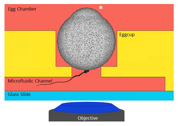
IVF in the microfluidic chip. The oocyte is maintained still in an eggcup equipped with a small opening at the bottom that communicates with a microfluidic channel. The star indicates the amicrovillar portion of the oocyte which is kept far from the opening on the channel. A selected acrosome reacted spermatozoon is introduced inside the channel and let free to interact with the restricted accessible membrane oocyte portion. The interaction zone is accurately imaged with a confocal microscope.
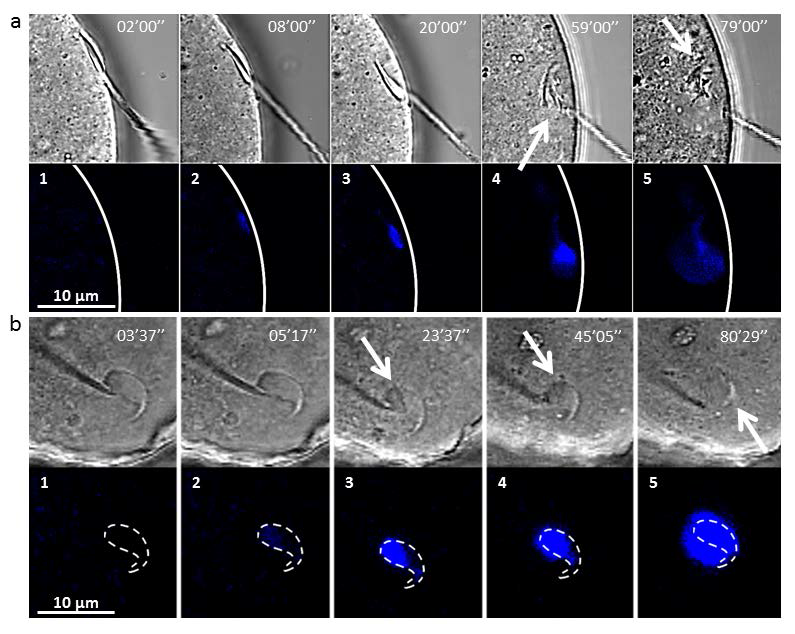
Snapshots of the sperm/egg interaction area during the fertilization process (bright field and fluorescence images, 405 nm wavelength). Timer indicates time after onset contact. Blue spots correspond to sperm DNA staining. (a)- Side view. a1-a3 Internalization and tilt of the spermatozoon head. a4-a5 sperm nucleus decondensation. Arrow on a4 indicates sperm membrane alteration. Arrow on a5 indicates the small portion of the spermatozoon head that remains intact after full decondensation. Flagellum seems to be stuck in the oocyte membrane. (b)- Front view with the microfluidic chip. The dashed line indicates the contour of the sperm head. b1-b3 Sperm DNA remains within these contours during 45 min. White arrow indicates sperm membrane alteration. b4-b5 the signal spreads out the contours which means that sperm DNA is decondensing. Arrow on b4 indicates sperm membrane alteration. Arrow on b5 indicates the small portion of the spermatozoon head that remains intact after full decondensation.