Eric Perez

Understanding the mechanical and adhesion properties of superficial skin cells
The protective function of biological surfaces that are exposed to the exterior of living organisms is the result of a complex arrangement and interaction of cellular components. This is the case of the most external cornified layer of skin, the stratum corneum (SC) made of corneocytes, the elementary ‘flat bricks’ and of a lipid matrix. These corneocytes are arranged in layers, and they adhere to each other through corneodesmosomes. Despite the well observed protective role of SC, the subtleties regarding the adhesion and mechanical interaction among the individual corneocytes are still poorly known. We have explored the interplanar (between layers) and peripheral (among cells within a layer) adhesions of single corneocytes by applying and measuring their detachment forces using glass microcantilevers. We have discovered key distinctive features: adhesion increases with depth in the SC; in the outermost layers, the population of corneocytes is not only heterogeneous, with strongly and weakly adhering corneocytes side by side, but corneocyte adhesion is anisotropic, stronger at the edges than on the flat sides. Each of these contributes to the mechanical integrity of the cutaneous barrier and therefore to the protective role of the SC.
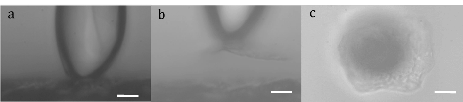
Cantilever glued to one corneocyte (a) before pulling, (b) after pulling and detaching one corneocyte, (c) front view of the detached corneocyte
Click here for a complete list of publications for E. Perez